An ultrasound scan — also called sonography — is one of the most widely used, versatile, and safest imaging tests in modern medicine. Using high-frequency sound waves instead of radiation, it creates real-time pictures of the organs, tissues, blood vessels, and even blood flow inside your body. It’s the same technology used to monitor a developing baby during pregnancy, but its applications extend far beyond obstetrics. To understand how ultrasound compares with other imaging methods, see our guide to radiology services at IPMC.
At Independent Physicians Medical Center (IPMC) in Northeast Philadelphia, we offer a comprehensive range of ultrasound services performed by experienced, registered sonographers and interpreted by board-certified radiologists — all in a private, comfortable outpatient setting.
How Does an Ultrasound Scan Work?
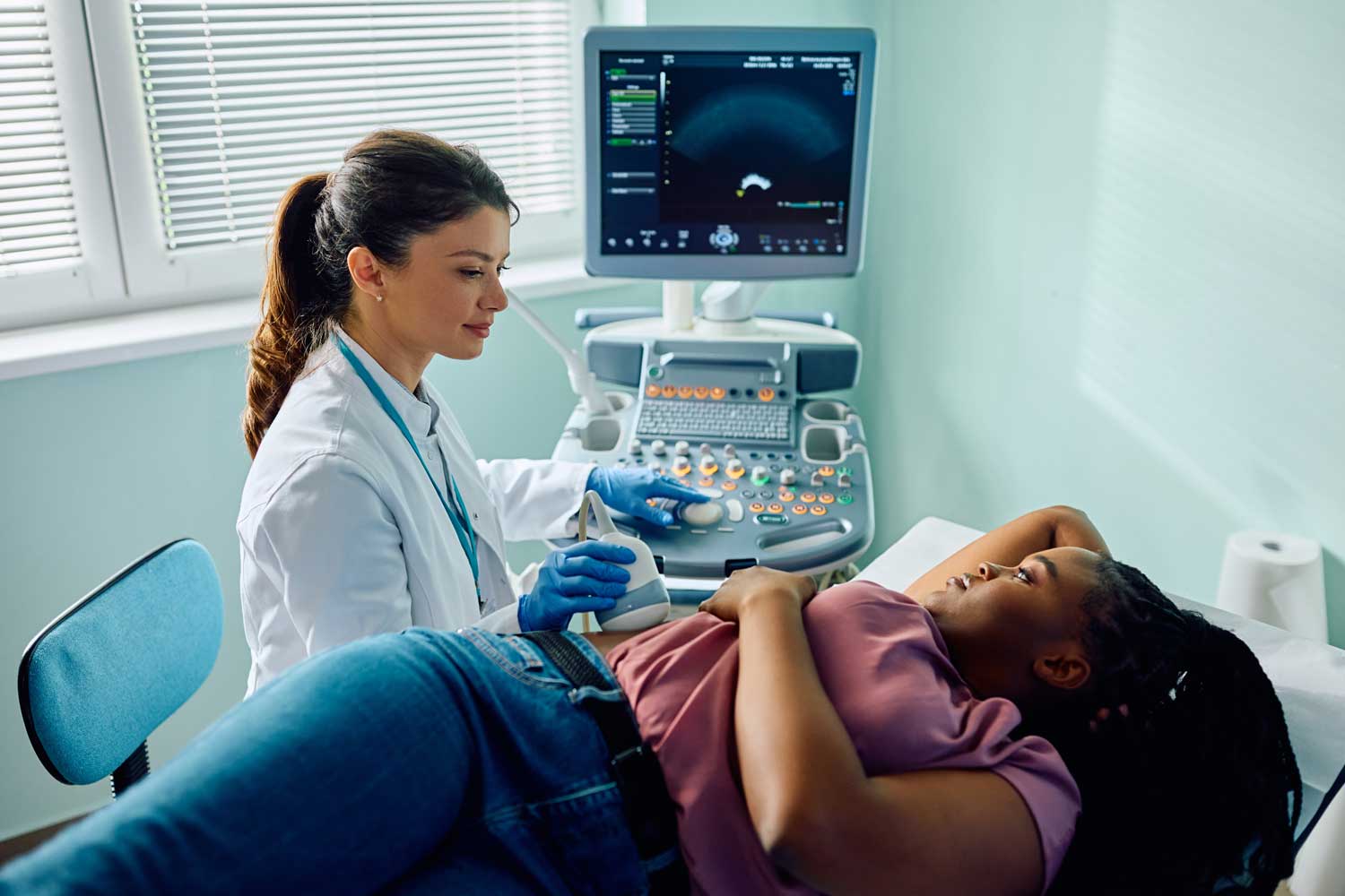
During an ultrasound exam, a small handheld device called a transducer is placed on your skin (or, in some cases, inserted into the body). The transducer emits high-frequency sound waves that travel into your body and bounce off internal structures — organs, tissues, fluid, and blood vessels. A computer captures these returning echoes and converts them into real-time images displayed on a monitor. According to RadiologyInfo.org — published jointly by the Radiological Society of North America (RSNA) and the American College of Radiology — ultrasound is a safe, well-established diagnostic tool with no known harmful effects at diagnostic levels.
Different tissues reflect sound waves differently. Fluid-filled structures like cysts appear dark (anechoic), solid organs appear in shades of gray, and dense structures like bone or calcifications appear bright white. This contrast allows the sonographer and radiologist to distinguish between normal and abnormal findings with remarkable precision.
Doppler ultrasound is a specialized technique that measures the speed and direction of blood flow through arteries and veins. Color Doppler adds color to the image to show flow direction, while spectral Doppler produces a waveform that reveals flow velocity. These techniques are invaluable for evaluating conditions like blood clots, narrowed arteries, and abnormal blood flow to organs.
What Can an Ultrasound Scan Be Used For?
Ultrasound has an extraordinarily wide range of clinical applications. See the full list of ultrasound types we offer at IPMC for more detail on each study.
- Abdominal imaging. Evaluating the liver, gallbladder, bile ducts, pancreas, spleen, kidneys, and abdominal aorta for conditions such as gallstones, fatty liver disease, kidney stones, cysts, tumors, and aneurysms. Learn more from the American College of Radiology’s guide to abdominal ultrasound.
- Pelvic imaging. Examining the uterus, ovaries, fallopian tubes, cervix, bladder, and prostate for fibroids, cysts, masses, endometriosis, and other conditions.
- Thyroid imaging. Evaluating thyroid nodules, goiter, and guiding fine needle aspiration biopsies. The American College of Radiology recommends ultrasound as the primary imaging tool for thyroid nodule evaluation.
- Breast imaging. Differentiating solid masses from fluid-filled cysts, evaluating mammographic findings, and guiding biopsies. Ultrasound is commonly used alongside mammography for a more complete picture of breast health.
- Vascular imaging. Assessing blood flow in the carotid arteries (stroke risk), leg veins (deep vein thrombosis), and other vessels.
- Musculoskeletal imaging. Evaluating tendons, ligaments, muscles, and joints for tears, inflammation, and fluid collections.
- Testicular imaging. Investigating lumps, pain, swelling, and blood flow abnormalities in the scrotum.
- Pregnancy monitoring. Tracking fetal development, estimating due dates, checking for complications, and guiding prenatal procedures.
- Procedure guidance. Directing needle placement during biopsies, fluid drainage, joint injections, and line insertions for greater accuracy and safety.
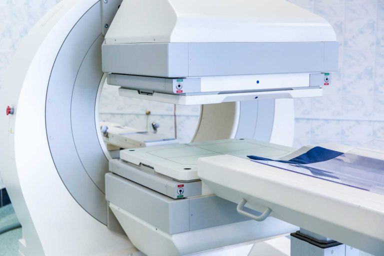
Because ultrasound uses no ionizing radiation, it is considered one of the safest imaging modalities and can be used repeatedly without cumulative risk — making it ideal for monitoring conditions over time and for imaging pregnant women, children, and patients of all ages. This is one important distinction from X-ray and CT scans, which use ionizing radiation. For a full comparison of imaging types, see our guide to MRI and CT scan vs. MRI comparison.
How to Prepare for Your Ultrasound Scan
Preparation varies by the type of exam. Our full ultrasound preparation guide has exam-specific instructions, but here is a general overview:
Abdominal Ultrasound
You will typically be asked to fast (no food or drink) for 8 to 12 hours before the exam. Fasting keeps the gallbladder distended with bile and reduces gas in the intestines, both of which improve image quality.
Pelvic Ultrasound
You may need to drink 32 ounces of water and arrive with a full bladder. A full bladder pushes the intestines out of the way and acts as a “window” that helps sound waves reach the pelvic organs.
Thyroid, Breast, Testicular, and Vascular Ultrasounds
Generally no preparation is needed. You can eat, drink, and take medications normally.
General Tips
Wear loose, comfortable clothing. You may be asked to change into a gown depending on the exam. Leave jewelry at home or be prepared to remove it. Our scheduling team will provide specific instructions when you book your appointment.
What Happens During an Ultrasound Scan at IPMC?
At IPMC, your exam will follow this general process:
- Check-in and positioning. You’ll be brought to a private exam room and positioned on an exam table — typically lying on your back, though you may be asked to turn onto your side for certain views.
- Applying the gel. The sonographer applies a clear, water-based gel to the area being examined. This gel eliminates air between the transducer and your skin (air blocks sound waves) and helps the transducer glide smoothly. The gel may feel slightly cool at first but warms quickly.
- Scanning. The sonographer presses the transducer gently against your skin and moves it across the area, capturing images from multiple angles. Real-time images appear on a monitor. You may see your organs on screen as the sonographer works. The sonographer may ask you to hold your breath, change positions, or take deep breaths at certain points to optimize the images.
- Doppler assessment (if applicable). If blood flow evaluation is part of your exam, you may hear a pulsing “whooshing” sound — this is the Doppler signal detecting blood moving through vessels.
- Duration. Most ultrasound exams take 20 to 45 minutes, though some specialized studies may take longer.
- Comfort. The exam is painless for most people. You may feel light pressure from the transducer, and certain areas (such as a tender gallbladder) may produce mild discomfort when pressed, but this is brief.
It’s important to know that sonographers are trained to capture high-quality diagnostic images, but they are not permitted to diagnose conditions or discuss results during the exam. Your images will be reviewed by a board-certified radiologist afterward.
After Your Ultrasound Scan
There is absolutely no recovery time. The gel is wiped off your skin, and you can eat, drink, drive, and return to all normal activities immediately. If you were asked to fast, you can eat right after.
A board-certified radiologist at IPMC will review all of the images and prepare a detailed report, which is sent to your referring doctor. Your doctor will contact you to discuss the results and any recommended follow-up — whether that’s reassurance that everything looks normal, additional imaging (such as an MRI or CT scan), a biopsy, or a referral to a specialist.
Ultrasound at IPMC
Why Choose IPMC for Your Ultrasound in Philadelphia
Expert Sonographers and Board-Certified Radiologists
Every ultrasound at IPMC is performed by a registered diagnostic sonographer and interpreted by a board-certified radiologist — with results sent directly to your referring physician.
Comprehensive Ultrasound Services
From abdominal and pelvic studies to vascular, thyroid, breast, and musculoskeletal imaging, we offer the full range of ultrasound types in one convenient location.
Convenient Location and Flexible Hours
Located at 9908 E. Roosevelt Blvd. in Northeast Philadelphia with onsite parking. Open Monday–Friday, 8AM–8PM — no hospital wait times.
Fast Appointments & Quick Results
Same-week scheduling and prompt results — so your care can move forward without delay. We accept most major insurances.
Schedule Your Ultrasound at IPMC
If your doctor has recommended an ultrasound to investigate symptoms or monitor a condition, Independent Physicians Medical Center is here to help with reliable, comfortable imaging close to home in Northeast Philadelphia.
- Call 215-464-3300 to schedule your ultrasound appointment.
- 9908 E. Roosevelt Blvd., Philadelphia, PA 19115
At IPMC, we believe medical imaging should be personal, efficient, and designed to support your health with confidence.