When it comes to breast cancer screening and early detection, technology continues to evolve — and 3D mammography, also known as tomosynthesis or digital breast tomosynthesis (DBT), is one of the most impactful advancements in recent years. This advanced imaging method provides clearer, more detailed views of breast tissue than traditional mammograms, helping doctors spot abnormalities earlier and with greater confidence.
At Independent Physicians Medical Center (IPMC) in Northeast Philadelphia, we offer advanced breast imaging services including 3D mammography as part of comprehensive breast health care, delivered in a welcoming outpatient setting. If you’re looking for 3D mammography in Philadelphia, our experienced team and state-of-the-art equipment are ready to help.
What Is 3D Mammography (Tomosynthesis)?
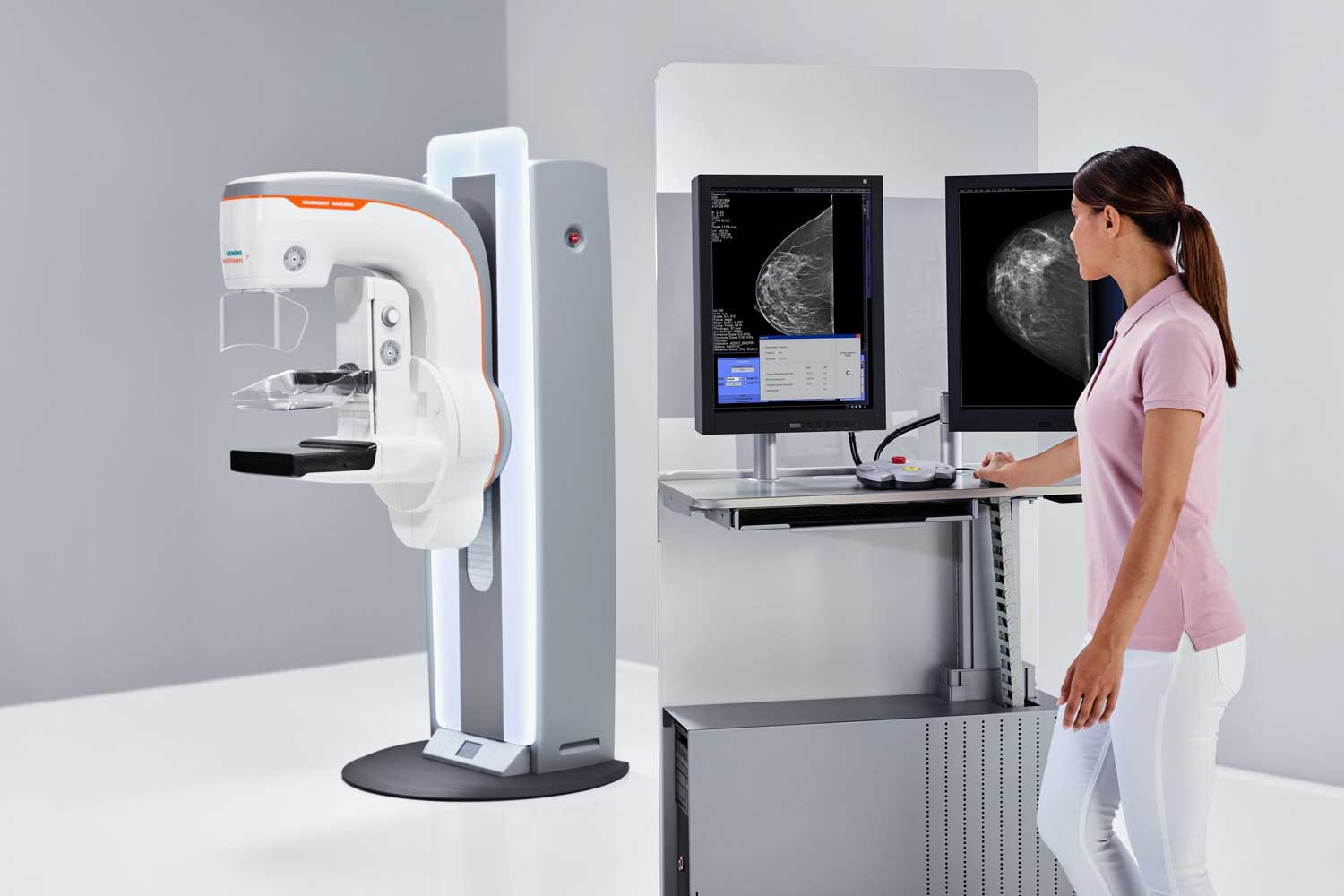
Tomosynthesis — also referred to as digital breast tomosynthesis (DBT) — is a specialized form of mammography that creates three-dimensional images of the breast. Instead of taking only one flat picture like a traditional mammogram, the imaging arm sweeps in a slight arc around the breast and captures multiple low-dose X-ray images from different angles. A computer then assembles these images into a detailed, layered 3D view that allows radiologists to examine breast tissue slice by slice.
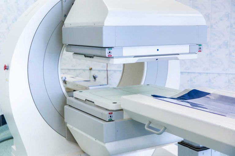
At IPMC, we use the MAMMOMAT Revelation (Siemens Healthineers) — a wide-angle tomosynthesis system that sweeps a full 50-degree arc around the breast, capturing 25 images per exam. Its wide-angle technology delivers the industry’s highest depth resolution, separating overlapping tissue layers for exceptional image clarity. It also provides automated breast density measurement at the point of examination — a valuable addition since high breast density is itself a risk factor for breast cancer.
According to the American College of Radiology, digital breast tomosynthesis is FDA-approved and increasingly recommended as part of routine breast cancer screening, particularly for women with dense breast tissue.
How Is 3D Mammography Different from a Traditional Mammogram?
Traditional 2D mammography takes a single flat image of the breast from two directions. When the breast is compressed, all the tissue overlaps in that one image — making it harder to distinguish between normal dense tissue and a potential tumor. Both can appear as white areas on a 2D image.
3D mammography solves this by capturing the breast in multiple thin “slices,” allowing the radiologist to scroll through the images layer by layer — similar to how a CT scan shows the body in cross-sections. This eliminates the confusion caused by overlapping tissue and reveals suspicious areas with much greater clarity.
For a broader comparison of imaging technologies, see our guide to what mammography is and how it works.
Key Benefits of 3D Mammography
Better Detection in Dense Breast Tissue
Dense breast tissue is common — many women have it — and it can make tumors harder to see on traditional mammograms because both dense tissue and cancer appear white on 2D images. Tomosynthesis helps separate overlapping structures, making suspicious areas far easier to identify even in dense breasts. The American Cancer Society notes that women with dense breasts have both a higher risk of breast cancer and a greater chance of having cancers missed by 2D mammograms.
Fewer Unnecessary Callbacks
Because of the 3D detail, tomosynthesis often reduces unnecessary callbacks for additional testing due to ambiguous findings on 2D images. Physicians can see more clearly whether a suspicious area is real or just overlapping tissue — reducing anxiety and follow-up imaging for patients.
Earlier Cancer Detection
Studies have shown that 3D mammography may detect more breast cancers — especially small or subtle lesions — compared with standard mammography alone. Earlier detection means more treatment options and better outcomes. The National Cancer Institute reports that finding breast cancer early, before symptoms appear, gives patients the greatest chance of successful treatment.
Low Radiation, High Detail
Despite capturing many more images than a standard mammogram, the MAMMOMAT Revelation system keeps radiation exposure minimal — at levels comparable to or slightly above a standard 2D mammogram. The added diagnostic detail comes without significant additional radiation risk.
Who Should Get a 3D Mammogram?
3D mammography is appropriate for most women undergoing routine breast cancer screening or diagnostic evaluation. It is especially recommended for:
- Women with dense breast tissue (categories C or D on a mammography report)
- Women at elevated risk for breast cancer due to family history, genetic mutations (BRCA1/BRCA2), or prior chest radiation
- Women with a previous abnormal mammogram requiring follow-up
- Women experiencing breast symptoms such as a lump, skin changes, or nipple discharge
- Women undergoing routine annual screening who want the most thorough evaluation available
Your doctor will determine whether 3D mammography, a standard mammogram, a breast MRI, or a combination provides the best assessment for your individual situation. For women at very high risk, breast MRI is often recommended alongside annual mammography.
What to Expect During a 3D Mammogram at IPMC
The 3D mammography experience is very similar to a traditional mammogram. Our full mammography preparation guide has everything you need to know before your visit.
- You’ll stand in front of the MAMMOMAT Revelation machine.
- A technologist positions one breast at a time on the imaging platform.
- The breast is gently compressed between two plates. The MAMMOMAT Revelation’s Personalized Soft Compression automatically adjusts to apply only the pressure your breast needs — making the exam more comfortable without compromising image quality.
- The imaging arm sweeps in a 50-degree arc around your breast, capturing 25 rapid low-dose X-ray images.
- A computer reconstructs these images into a detailed 3D view — slice by slice — for the radiologist to review.
- The process is quick. The entire exam typically takes about 20 minutes, and there is no recovery time.
On the day of your mammogram, avoid wearing deodorant, perfume, or lotion on your torso — these can interfere with image quality. Schedule your exam when your breasts are least likely to be tender (usually the week after your period).
Mammography at IPMC
Why Choose IPMC for 3D Mammography in Philadelphia?
Convenient Location and Flexible Hours
Easily accessible with onsite parking. Open Monday–Friday from 8AM to 8PM to fit your schedule.
Advanced Mammography Technology
Personalized, Outpatient Breast Imaging
Fast Appointments & Quick Results
Schedule Your 3D Mammogram at IPMC
Early detection saves lives. If you’re due for a screening mammogram or have been advised to undergo 3D mammography in Philadelphia, IPMC makes it easy — same-week appointments, advanced wide-angle tomosynthesis, and results delivered directly to your doctor.
- Call 215-464-3300 to schedule your appointment.
- Visit us at 9908 E. Roosevelt Blvd., Philadelphia, PA 19115.
At Independent Physicians Medical Center, we believe breast cancer screening should be accurate, comfortable, and tailored to you — helping you and your doctor make confident decisions about your health.