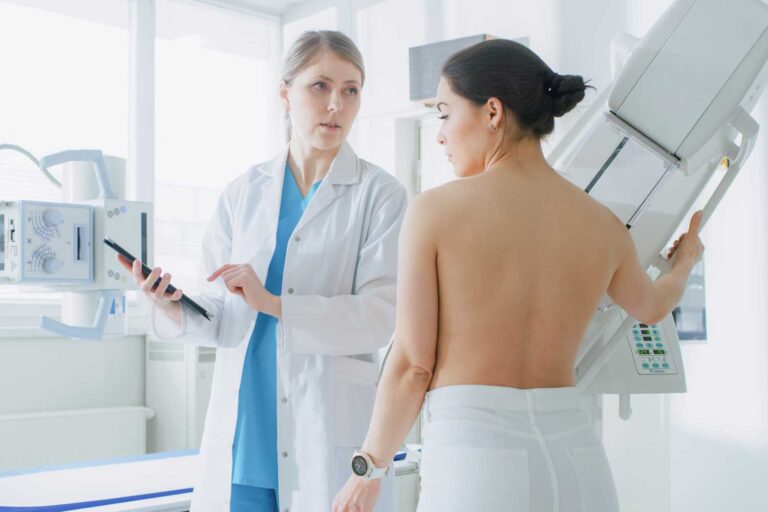
What Is a Breast Ultrasound?
A breast ultrasound (also called breast sonography) uses a handheld device called a transducer to emit sound waves that bounce off internal structures and create detailed images of the breast. These images are called sonograms and show how breast tissue and any abnormalities appear in real time.
Unlike X-ray-based mammograms, ultrasound does not use radiation, making it a safe option for many patients, including those who are pregnant or require repeated imaging.
Why Your Doctor May Order a Breast Ultrasound
Breast ultrasound is often recommended for a variety of reasons:
Investigating Abnormalities
If a lump, pain, skin change, or unusual nipple discharge is detected during a physical exam, your doctor may use ultrasound to get a closer look.
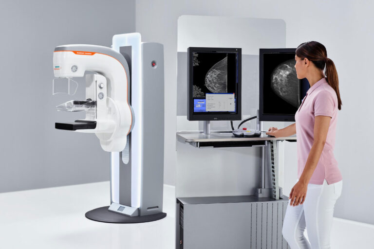
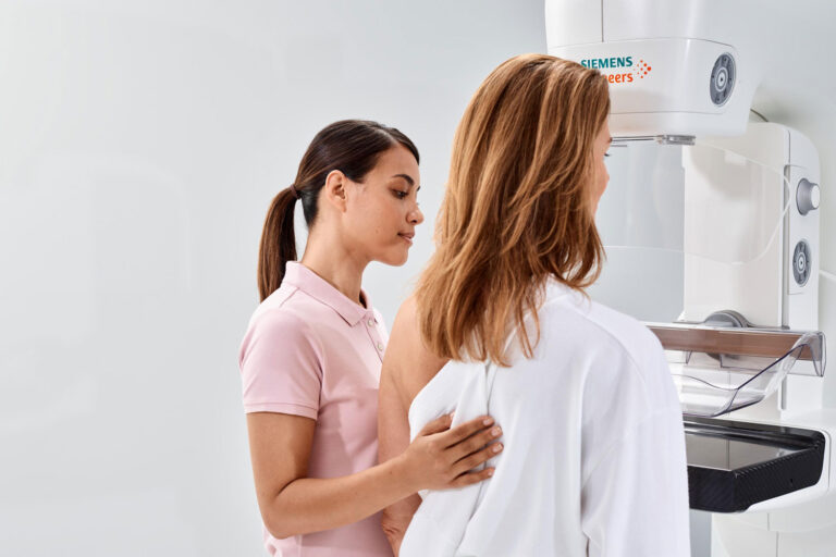
Following Up After a Mammogram
If a mammogram shows an area of concern — especially in denser breast tissue — ultrasound helps distinguish between fluid-filled cysts and solid masses that might require further evaluation.
Complementing Other Imaging
Ultrasound is particularly helpful for women with dense breasts, where a mammogram may be less sensitive. It may be used in addition to mammography to provide more detailed imaging.
Guiding Procedures
Ultrasound can also assist in procedures like biopsies or cyst drainage by helping guide needles precisely to the area of interest.
Special Situations
For patients who should avoid radiation (such as during pregnancy), ultrasound offers a safe alternative for imaging breast tissue.
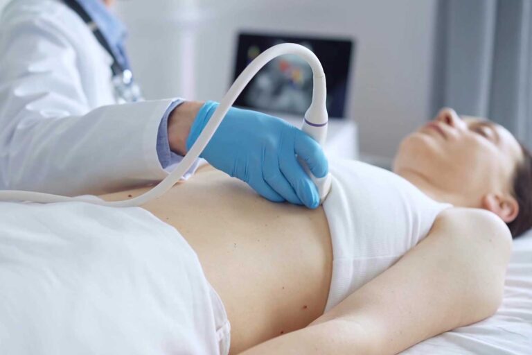
What to Expect During the Test
A breast ultrasound is typically straightforward and comfortable:
- You’ll be asked to remove clothing from the waist up and lie on an exam table.
- A clear gel is applied to your breast to help transmit sound waves.
- A sonographer moves the transducer over your breast to capture images.
- The test typically takes about 20–30 minutes.
There’s no radiation exposure, and most patients can resume normal activities immediately afterward.
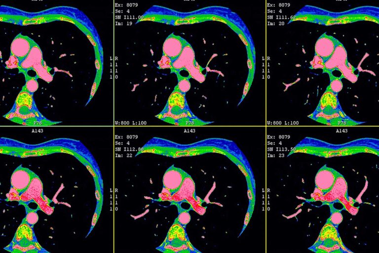
What Ultrasound Can Show
Breast ultrasound can help determine:
- Whether a breast lump is fluid-filled (a cyst) — usually benign — or solid, which might need further testing.
- The location, size, and shape of abnormalities.
- Whether additional imaging or a biopsy may be needed.
While ultrasound is an excellent tool for evaluating specific areas, it does not replace mammography as the primary screening test for breast cancer. Mammograms remain the standard for routine screening in women at average risk.
Benefits of Breast Ultrasound
Breast ultrasound offers several advantages:
- Safe and non-invasive — no radiation exposure.
- Useful in dense breast tissue — provides additional detail where mammograms may be less clear.
- Real-time imaging — helpful in guiding biopsies and procedures.
- Comfortable — most patients find it painless and quick.
Ultrasound at IPMC
Why Choose IPMC for Your Ultrasound in Philadelphia
Advanced Ultrasound Technology
Convenient Location and Flexible Hours
Easily accessible with onsite parking. Open Monday–Friday from 8AM to 8PM to fit your schedule.
Comfortable Outpatient Experience
Fast Appointments & Quick Results
Schedule Your Breast Ultrasound at IPMC
If your doctor has recommended a breast ultrasound to evaluate symptoms, follow up on a mammogram, or guide a procedure, Independent Physicians Medical Center is here to help with dependable, compassionate care in Northeast Philadelphia.
- Call 215-464-3300 to schedule your appointment.
- 9908 E. Roosevelt Blvd., Philadelphia, PA 19115
At IPMC, we believe imaging should be personal, efficient, and tailored to your health needs — giving you clarity and confidence in your care.