
If you or a loved one is facing a cancer concern, you may wonder whether an MRI can help detect or evaluate the disease. The answer is yes — MRI is one of the most powerful and versatile imaging tools in cancer care. Its exceptional ability to produce detailed images of soft tissues makes it invaluable for finding tumors, determining their extent, guiding treatment decisions, and monitoring how well therapy is working.
At Independent Physicians Medical Center (IPMC) in Northeast Philadelphia, we offer advanced MRI technology that plays a critical role in cancer detection and care, all in a comfortable outpatient setting.
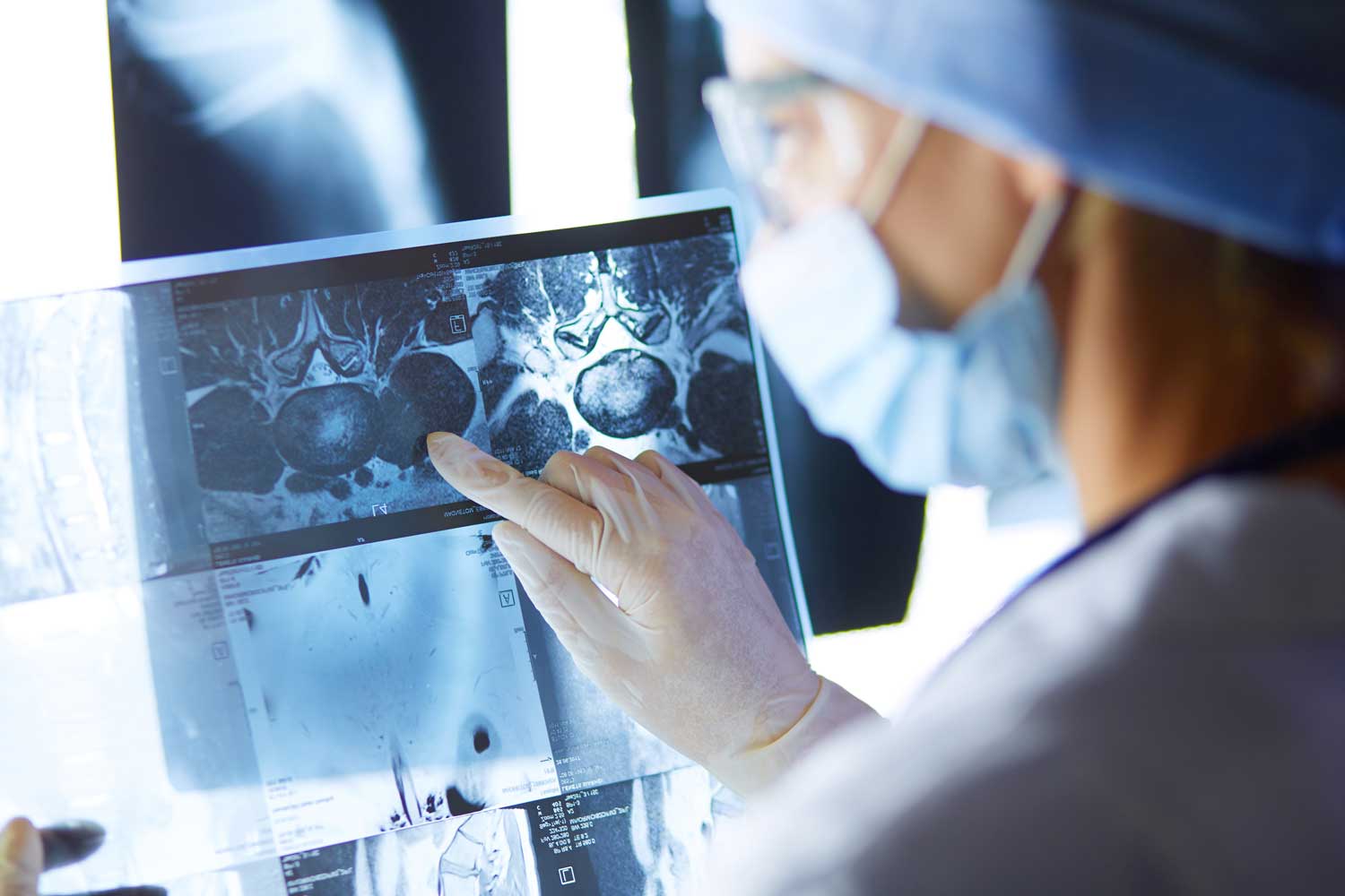
How Does MRI Help Detect Cancer?
MRI uses strong magnetic fields and radio waves to create highly detailed images of organs and soft tissues throughout the body. Because cancer typically affects soft tissues, MRI is exceptionally effective at identifying tumors that other imaging methods — such as X-rays or even CT scans — may miss.
MRI can detect cancer by identifying abnormal masses or areas of tissue that differ from their surroundings in size, shape, or signal characteristics. When gadolinium contrast dye is used, cancerous areas often enhance (light up) differently than normal tissue because tumors typically develop their own blood supply (a process called angiogenesis). The pattern of contrast enhancement provides important clues about whether a mass is likely benign or malignant.
Advanced MRI techniques such as diffusion-weighted imaging (DWI) can also detect cancers by measuring how water molecules move within tissues. Cancer cells tend to be densely packed, restricting the movement of water, which produces a characteristic bright signal on DWI that helps radiologists identify suspicious areas.
What Types of Cancer Can MRI Help Detect?
MRI is used to evaluate many types of cancer, including:
- Brain and spinal cord tumors — MRI is the primary imaging modality for brain cancer detection and is far superior to CT for this purpose
- Breast cancer — MRI is the most sensitive imaging test for breast cancer and is especially valuable for high-risk screening, staging newly diagnosed cancers, and evaluating dense breast tissue
- Prostate cancer — multiparametric MRI (mpMRI) has revolutionized prostate cancer detection and is now a key tool in diagnosis, biopsy guidance, and active surveillance
- Liver cancer and liver metastases — MRI with liver-specific contrast agents provides the highest sensitivity for detecting small liver tumors
- Cervical, uterine, and ovarian cancers — MRI is essential for staging pelvic gynecological cancers
- Rectal cancer — MRI is the gold standard for local staging of rectal cancer, helping surgeons plan the best approach
- Kidney cancer — MRI can characterize kidney masses and is an alternative to CT for patients who cannot receive CT contrast
- Bone and soft tissue sarcomas — MRI provides superior visualization of these tumors and their relationship to surrounding muscles, nerves, and blood vessels
- Head and neck cancers — MRI is valuable for assessing tumor extent and involvement of adjacent structures
- Pancreatic cancer — MRI and MRCP complement CT in evaluating pancreatic tumors, especially those involving the bile ducts
MRI is also widely used for cancer staging (determining the size, location, and extent of the cancer), monitoring treatment response (showing whether a tumor is shrinking, stable, or growing during chemotherapy or other therapies), and detecting cancer recurrence after treatment.
MRI vs. Other Imaging for Cancer
While CT scans, PET scans, ultrasound, and X-rays each play important roles in cancer care, MRI offers distinct advantages in many situations. Its superior soft tissue contrast makes it the preferred choice for imaging the brain, breast, prostate, liver, pelvis, and musculoskeletal system. It also does not use ionizing radiation, which is beneficial for patients who need repeated scans over the course of treatment — including children and young adults.
Your doctor may use MRI in combination with other tests to build the most complete picture of your condition. For example, a CT scan may be used for initial detection, and an MRI may follow for more detailed characterization. PET/MRI is an emerging combined technique that provides both metabolic and structural information in a single exam.
What to Expect and Follow-Up
The procedure is the same as a standard MRI. You’ll lie on a padded table that slides into the machine, and the scan typically takes 30 to 60 minutes depending on the area being examined. Contrast dye is commonly used and will be given through an IV. A technologist communicates with you via intercom throughout.
There is no downtime after the scan. A board-certified radiologist at IPMC will interpret the images and provide a detailed report to your referring physician or oncologist. The results will help guide your treatment plan and next steps — whether that’s additional testing, a biopsy, treatment initiation, or reassurance that the scan is normal.
MRI at IPMC
Why Choose IPMC for MRI in Northeast Philadelphia
Convenient Location and Flexible Hours
Easily accessible with onsite parking. Open Monday–Friday from 8AM to 8PM to fit your schedule.
Advanced MRI Technology
Our equipment delivers detailed images that help your doctor make accurate diagnoses and guide treatment.
Comfortable Outpatient Setting
Fast Appointments & Quick Results
Schedule Your MRI at IPMC
If your physician has recommended an MRI to evaluate a suspicious finding, monitor a known condition, or aid in cancer care planning, Independent Physicians Medical Center is here to help.
- Call 215-464-3300 to schedule your appointment.
- 9908 E. Roosevelt Blvd., Philadelphia, PA 19115
- Monday–Friday, 8AM–8PM
At IPMC, advanced imaging and compassionate care help you and your doctor get the information necessary for confident diagnosis and treatment planning.


