An X-ray is the oldest, fastest, and most widely used diagnostic imaging test in medicine. In seconds, it produces detailed images of the bones, joints, lungs, and other internal structures — giving your doctor the information needed to diagnose fractures, infections, lung conditions, and more without surgery or complex preparation.
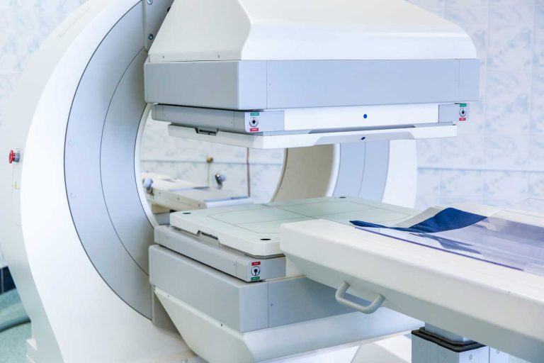
If your doctor has ordered an X-ray in Philadelphia, Independent Physicians Medical Center (IPMC) in Northeast Philadelphia provides advanced digital X-ray imaging using the Siemens Multix system — a state-of-the-art digital radiography platform that delivers sharp images with minimal radiation exposure, in a comfortable outpatient setting with no hospital wait times.
What Is an X-Ray and How Does It Work?
An X-ray uses a small, controlled beam of ionizing radiation to pass through your body and project an image onto a detector (or, in older systems, photographic film). Different tissues absorb the X-ray beam at different rates:
- Dense structures like bone absorb most of the radiation and appear bright white on the image.
- Soft tissues like muscle and fat absorb less and appear in shades of gray.
- Air-filled spaces (such as the lungs) absorb almost none and appear dark or black.
This contrast makes X-rays exceptionally good at showing bone structure and detecting abnormalities in the chest and abdomen — and because the scan takes only seconds and requires no preparation for most studies, it is typically the first imaging test ordered in many clinical situations.
IPMC uses digital radiography rather than traditional film. Digital X-ray images are captured electronically and immediately available for review on a monitor, enabling faster diagnosis, lower radiation doses, and the ability to digitally adjust image contrast and brightness to optimize diagnostic detail.
According to RadiologyInfo.org (published by the American College of Radiology), X-rays have no immediate side effects in the typical diagnostic range, and the benefit of an accurate diagnosis far outweighs the small radiation risk for most patients.
What Can an X-Ray Diagnose? Common Uses by Body Area
X-rays are used across a wide range of clinical situations. The table below covers the most common applications and what each type of X-ray can show.
| Type of X-ray | What it evaluates | Common conditions detected |
|---|---|---|
| Chest X-ray | Lungs, heart size, major blood vessels, ribs, and diaphragm | Pneumonia, COPD, heart enlargement, pleural effusion, rib fractures, lung masses. See our chest X-ray guide. |
| Bone / extremity X-ray | Arms, legs, hands, feet, wrists, ankles | Fractures, dislocations, bone tumors, growth plate injuries in children |
| Spine X-ray | Cervical, thoracic, or lumbar vertebrae and disc spaces | Compression fractures, scoliosis, spondylosis, spinal alignment, degenerative disc disease |
| Joint X-ray (hip, knee, shoulder) | Bone surfaces, joint space, and alignment | Osteoarthritis, joint space narrowing, bone spurs, joint deformity. See our X-ray for knee osteoarthritis guide. |
| Abdominal X-ray (KUB) | Kidneys, ureters, bladder, bowel gas pattern | Bowel obstruction, calcified kidney stones, free air (bowel perforation), constipation evaluation. See our X-ray for kidney stones guide. |
| Sinus X-ray | Frontal, maxillary, ethmoid, and sphenoid sinuses | Sinusitis (air-fluid levels), sinus opacification, polyps. See our sinus X-ray guide. |
| Skull X-ray | Skull bones and sutures | Skull fractures, bone lesions; largely replaced by CT for head trauma |
| Foreign body X-ray | Any body region where a foreign object is suspected | Swallowed or inhaled objects, retained surgical hardware, metallic foreign bodies in soft tissue |
X-Ray vs. CT Scan vs. MRI: Which Does Your Doctor Choose?
X-ray, CT, and MRI each have distinct strengths. Your doctor selects the right test based on what they need to see. The table below summarizes the key differences. For a more detailed comparison, see our full guide: CT Scan vs. MRI: What’s the Difference?
| X-ray | CT scan | MRI scan | |
|---|---|---|---|
| Radiation | Very low | Low–moderate | None |
| Scan time | Seconds | 5–15 min | 20–60 min |
| Bone detail | Excellent | Excellent | Good |
| Soft tissue detail | Limited | Good | Excellent |
| Chest / lungs | First choice | More detail | Limited use |
| Fractures | First choice | Better for complex / subtle | Limited use |
| Brain, spine, joints | Limited | Good for bone | First choice |
| Cost (general) | Lowest | Moderate | Highest |
| Best for emergencies | Yes — fastest | Yes — comprehensive | Too slow for acute |
Is an X-Ray Safe? Understanding Radiation Exposure
Radiation is one of the most common concerns patients have about X-rays. The context helps: X-ray radiation doses used in diagnostic imaging are very small, and the risk from a single study is extremely low for most patients.
| Study or exposure source | Approximate effective dose | Equivalent natural background radiation |
|---|---|---|
| Chest X-ray (PA) | 0.02 mSv | About 2.4 days of background radiation |
| Hand or foot X-ray | 0.001 mSv | A few hours of background radiation |
| Lumbar spine X-ray | 1.5 mSv | About 6 months of background radiation |
| Abdominal X-ray | 0.7 mSv | About 2.5 months of background radiation |
| Abdomen / pelvis CT scan | 8–10 mSv | About 2.5–3 years of background radiation |
| Annual natural background radiation (US average) | ~3 mSv/year | Baseline — from soil, cosmic rays, radon, food |
Key safety points:
- For most adults, the diagnostic benefit of a well-indicated X-ray far outweighs the very small radiation risk.
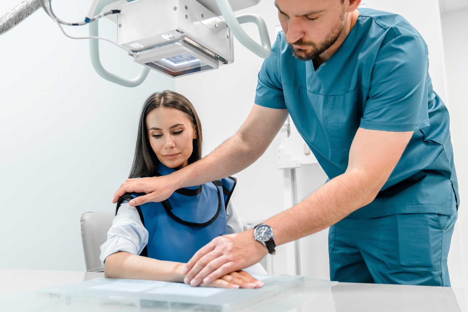
- Pregnant women should always inform the technologist and their doctor before an X-ray. When imaging is medically necessary, lead shielding is used to protect the abdomen. Many X-ray studies — particularly of the extremities, chest, and head — involve minimal scatter to the uterus.
- Children are more sensitive to radiation than adults. IPMC technologists use pediatric-appropriate protocols and the lowest dose that still produces a diagnostic image.
- The U.S. Food and Drug Administration notes that medical X-ray imaging provides benefits that generally far exceed the risks when used appropriately — and recommends that patients discuss any concerns with their doctor before declining a recommended study.
- If your doctor believes you need repeated imaging or requires detailed soft tissue evaluation without radiation, they may recommend an MRI or ultrasound instead.
How to Prepare for Your X-Ray
X-rays require less preparation than any other imaging test. Our full X-ray preparation guide covers exam-specific instructions. Here are the key points:
| Situation | Preparation needed |
|---|---|
| Most X-rays (extremities, chest, spine, joints, skull, sinuses) | ✓ None. You can eat, drink, and take medications normally. Arrive and go. |
| KUB or abdominal X-ray | If you have recently had a CT scan with contrast dye, barium swallow, or barium enema, wait at least 10 days before your abdominal X-ray. Residual contrast material can obscure findings. Notify IPMC when booking. |
| Clothing and jewelry | Leave jewelry at home. Remove piercings, watches, and any metal accessories from the area being imaged. Wear loose, comfortable clothing — you may be asked to change into a gown for certain studies. |
| Pregnancy | Always inform the technologist and your doctor if you are or may be pregnant before any X-ray, even one involving the extremities. Your care team will determine whether the exam is safe to proceed and use appropriate shielding. |
| Prior imaging | If you have previous X-rays of the same area from another facility, bring them or make them available. Comparing current images to prior ones can significantly improve diagnostic accuracy. |
What Happens During an X-Ray at IPMC?
- Positioning. A technologist positions you in front of the digital detector panel. Depending on the study, you may stand, sit, or lie on a table. The Siemens Multix system can accommodate patients in all positions — including those with mobility limitations.
- Holding still. You’ll be asked to stay completely still for a fraction of a second while the image is taken. Movement blurs the image. For chest X-rays, you may be asked to take a deep breath and hold it briefly.
- Multiple views. Most X-ray studies require two or more views taken from different angles — for example, front (AP or PA) and side (lateral) projections — to give the radiologist a complete picture of the anatomy.
- Duration. The entire exam takes just 5 to 15 minutes, including positioning and taking multiple views. The actual radiation exposure during each image lasts only a fraction of a second.
- After the exam. There is no recovery time. You can eat, drive, and return to all normal activities immediately.
After Your X-Ray: Results and Next Steps
After your X-ray, a board-certified radiologist at IPMC reviews every image and prepares a detailed report for your referring physician. For outpatient studies, results are typically available within 24 to 48 hours and sent directly to your doctor.
Depending on what the X-ray shows, your doctor may:
- Reassure you that nothing significant was found
- Recommend a follow-up X-ray at an interval to monitor healing or progression (e.g., fracture healing, scoliosis monitoring)
- Order additional imaging for a more detailed view — such as a CT scan for complex fractures or suspected internal bleeding, an MRI for soft tissue injuries, or an ultrasound for fluid collections or organ evaluation
- Refer you to a specialist based on the findings
- Begin or adjust treatment based on what the X-ray reveals
X-Ray at IPMC
Why Choose IPMC for Your X-Ray in Philadelphia?
Siemens Multix Digital Radiography
Board-Certified Radiologists
Fast, No-Wait Imaging
Convenient Location and Flexible Hours
Located at 9908 E. Roosevelt Blvd. in Northeast Philadelphia with onsite parking. Open Monday–Friday, 8AM–8PM. We accept most major insurance plans.
Schedule Your X-Ray at IPMC in Philadelphia
If your doctor has recommended an X-ray, IPMC provides fast, digital imaging in a comfortable outpatient setting in Northeast Philadelphia — with results delivered directly to your physician.
- Call 215-464-3300 to schedule your appointment.
- 9908 E. Roosevelt Blvd., Philadelphia, PA 19115
- Monday–Friday, 8AM–8PM
At Independent Physicians Medical Center, we believe medical care should be personal, efficient, and focused on your peace of mind—starting with your imaging experience.