Understanding Nuclear Medicine
What is Nuclear Medicine?
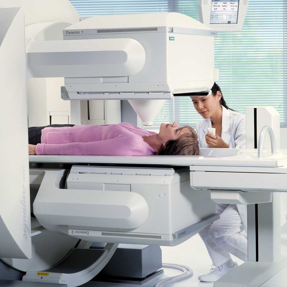
Unlike traditional radiology, which captures images using external radiation sources (like X-rays or CT scans), nuclear medicine uses small amounts of radiopharmaceuticals—radioactive substances introduced into the body to highlight specific organs or cellular activity. Once administered (usually orally or intravenously), these tracers travel to the target area, where a specialized gamma camera captures real-time images of the body’s internal function.
Rather than focusing only on anatomy, nuclear medicine shows how organs are working, making it ideal for detecting early-stage diseases such as thyroid disorders, heart conditions, bone abnormalities, and even metastatic cancer.
Benefits of Nuclear Medicine
The Diagnostic Advantage
Nuclear medicine offers a powerful way to see how the body is functioning from the inside out. This innovative imaging technique provides unique benefits that go beyond traditional scans—making it a valuable tool for early diagnosis, treatment planning, and monitoring a wide range of health conditions.
Early Detection of Disease – Nuclear medicine can reveal signs of illness before structural changes appear on other imaging tests.
Functional Insight – It focuses on physiology, helping doctors understand how well an organ or system is functioning—not just how it looks.
Minimally Invasive – Most procedures involve just a small injection or oral dose of a tracer, with no incisions or recovery time.
Safe and Effective – While it involves a small amount of radiation, nuclear medicine procedures are generally safe and carefully controlled. The benefits in diagnostic accuracy often far outweigh the minimal risk.
Versatile Diagnostic Tool – Useful in evaluating conditions in the heart, bones, lungs, kidneys, thyroid, liver, and more.


Imaging Excellence
Advanced Functional Imaging for Accurate Diagnoses
At IPMC Medical Center in Northeast Philadelphia, we’re proud to offer comprehensive nuclear medicine services—a highly specialized branch of diagnostic imaging that helps physicians evaluate organ function and detect diseases at their earliest stages. Using state-of-the-art technology and a patient-centered approach, we provide fast, accurate results in a comfortable outpatient setting.
Common Nuclear Medicine Tests We Offer:
- Bone Scans
- Thyroid Scans
- Renal (Kidney) Scans
- Hepatobiliary (HIDA) Scans
- Gastric Emptying Studies
- Parathyroid Scans
- MUGA (Cardiac Function) Scans
- Nuclear Stress Tests
Trusted Specialists
Nuclear Medicine at IPMC
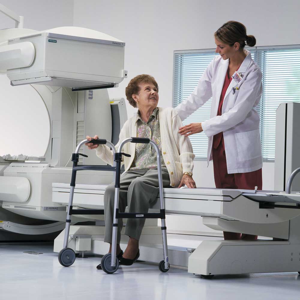
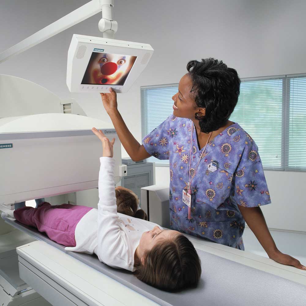
At IPMC, we perform all nuclear medicine tests on the Siemens E.Cam Duel gamma camera—a leading-edge system known for its clarity, speed, and open design. This setup is ideal for patients who are claustrophobic, use wheelchairs, or require stretchers. Our board-certified radiologists and experienced technologists ensure precise image capture and interpretation in a comfortable environment.
Whether you’re referred for routine testing or a specialized scan, our team at IPMC ensures a smooth, supportive experience from check-in to results.
Why Choose IPMC for Nuclear Medicine in Philadelphia?
- Convenient Northeast Philadelphia location
- Same-day and next-day appointments available
- On-site testing with fast turnaround times
- Compassionate care from experienced professionals
- Advanced imaging using trusted Siemens technology

