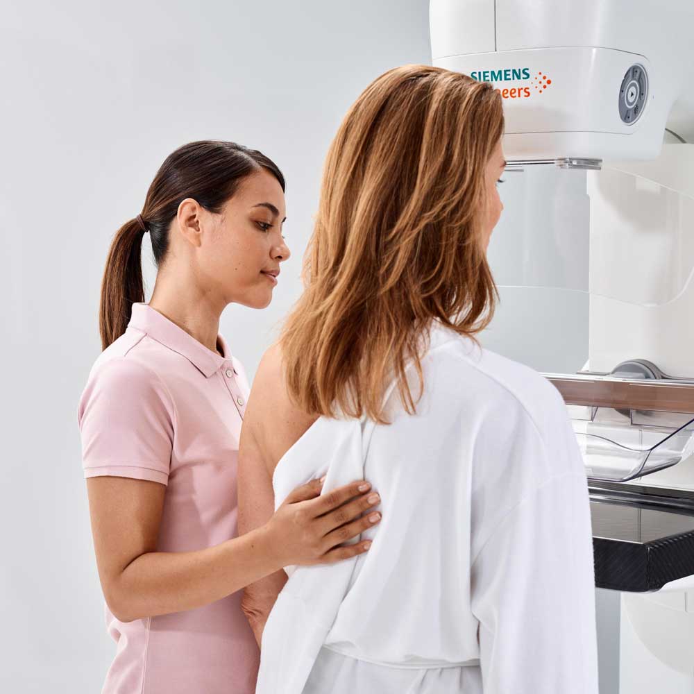
Understanding Mammography
What is Mammography?
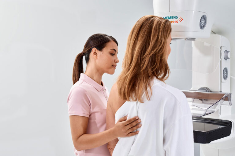
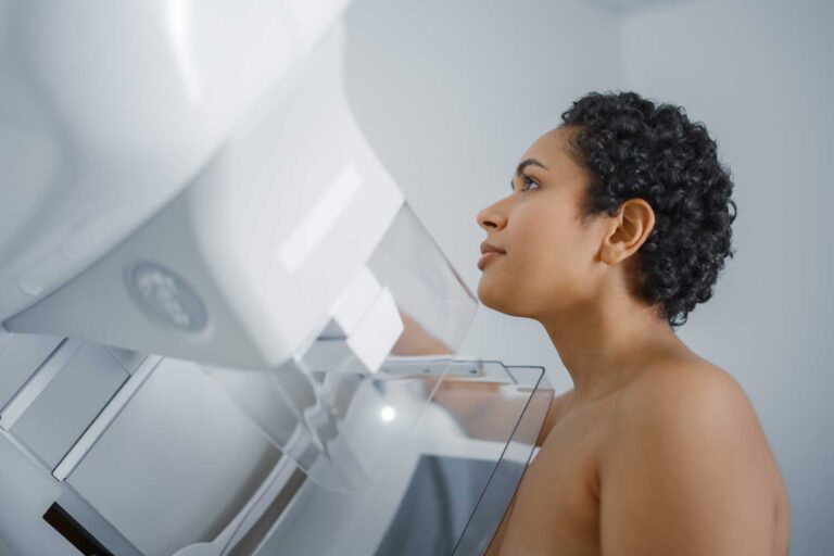
Mammography is a specialized medical imaging technique that uses low-dose X-rays to examine breast tissue. It’s the most effective screening tool for the early detection of breast cancer, often identifying changes in the breast before physical symptoms develop. There are two types of mammography: screening mammography, which is routine and preventive, and diagnostic mammography, which investigates specific concerns such as lumps, pain, or unusual changes.
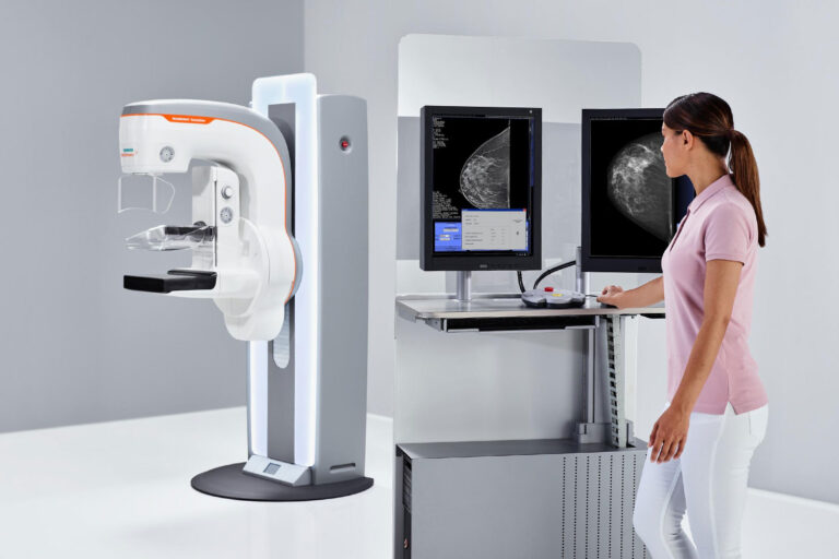
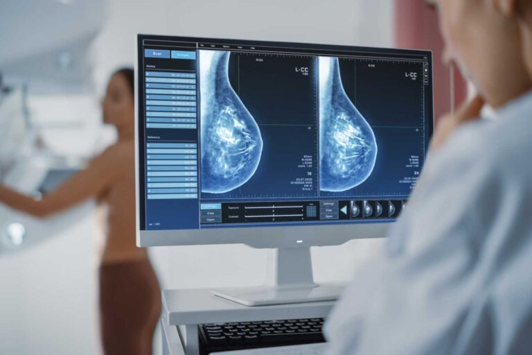
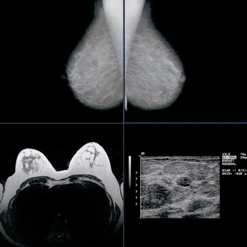
At IPMC in Northeast Philadelphia, we use advanced imaging systems including 3D mammography (Digital Breast Tomosynthesis) for a more detailed, accurate view of breast tissue. This technology allows our radiologists to detect abnormalities with greater precision—especially in patients with dense breast tissue.
Benefits of Mammography
Why Mammography Matters
Mammography offers powerful advantages in the early detection and diagnosis of breast conditions—often before symptoms appear.
Early Detection Saves Lives — Detecting breast cancer at an early stage dramatically improves treatment outcomes and survival rates.
High Accuracy with 3D Technology — Our 3D mammograms provide clearer images, reducing the chance of false positives and unnecessary follow-up tests.
Safe and Low Radiation — Mammography uses very low levels of radiation and is considered safe for regular screening.
Fast and Non-Invasive — The procedure typically takes 20 minutes and requires no recovery time.
Recommended for Routine Screening — The American Cancer Society recommends yearly screenings starting at age 40 for women at average risk.
Imaging Excellence
Mammography at IPMC
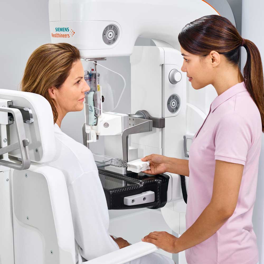
At Independent Physician Medical Center (IPMC), your comfort and care come first. Our state-of-the-art imaging center is equipped with a Siemens Healthineers MAMMOMAT Revelation mammography system, delivering exceptional image quality while keeping radiation exposure to a minimum. Our experienced radiology team ensures a smooth, respectful experience from check-in to results.
Located conveniently in Northeast Philadelphia, IPMC offers easy appointment scheduling, minimal wait times, and same-day availability for many imaging services. Whether you need a routine screening or a follow-up diagnostic exam, you can count on us for timely, compassionate care.
Simply fill out the form, and our team will contact you to confirm your appointment.
From News & Resources
Mammography Insights and Patient Resources
December 28, 2024
June 4, 2024
August 4, 2024
March 7, 2025
January 28, 2025
September 4, 2016
