
At Independent Physicians Medical Center (IPMC) in Northeast Philadelphia, we offer advanced knee MRI imaging in a relaxed outpatient setting with experienced technologists and board-certified radiologists who specialize in musculoskeletal imaging.
What Is a Knee MRI?
A knee MRI uses strong magnets and radio waves to produce highly detailed images of every structure inside and around your knee joint — including bones, cartilage, meniscus, ligaments (such as the ACL, PCL, MCL, and LCL), tendons, muscles, and the joint capsule (synovium). Unlike X-rays, which only reveal bones, an MRI shows soft tissue with remarkable clarity, making it the most important imaging tool for evaluating most knee problems.
The knee is a complex hinge joint made up of three bones (the femur, tibia, and patella), two crescent-shaped cartilage pads (the medial and lateral meniscus), four major ligaments, multiple tendons, and fluid-filled bursae. Because so many structures are packed into a relatively small space, an MRI’s ability to distinguish between them is invaluable.
Why Would Your Doctor Order a Knee MRI?
Your physician may recommend a knee MRI for:
- Unexplained or persistent knee pain that hasn’t responded to rest and basic treatment
- Swelling that doesn’t improve or keeps coming back
- A knee that gives out, locks, or catches during movement — these symptoms often suggest a meniscus tear or loose body in the joint
- A suspected torn meniscus, which is extremely common in both athletes and older adults
- A suspected ligament tear, particularly the ACL (anterior cruciate ligament) or MCL (medial collateral ligament) — common after sports injuries or twisting motions
- Cartilage damage, including chondromalacia (softening of the kneecap cartilage) and osteochondral defects
- Arthritis evaluation — MRI can show early cartilage loss that isn’t yet visible on X-rays
- Patellar tracking problems or kneecap instability
- Baker’s cyst (a fluid-filled cyst behind the knee)
- Bone bruises, stress fractures, or fractures not visible on X-ray
- Tendon problems, such as patellar tendinitis (“jumper’s knee”) or quadriceps tendon tears
- Pre-surgical evaluation before arthroscopy, knee replacement, or ligament reconstruction
- Monitoring progress after knee surgery or treatment
How to Prepare for Your Knee MRI
Preparation is minimal. You can eat, drink, and take your medications normally. Wear comfortable clothing — sweatpants and a t-shirt without metal are ideal. Remove any knee braces, jewelry, or metal objects. If you have surgical hardware in your knee (screws, plates, or an artificial joint), tell the technologist — most modern orthopedic implants are MRI-safe, but it’s always verified. Let your doctor know if you’re claustrophobic, though for a knee MRI, only your lower body enters the scanner, which many patients find much more tolerable.
What Happens During a Knee MRI?
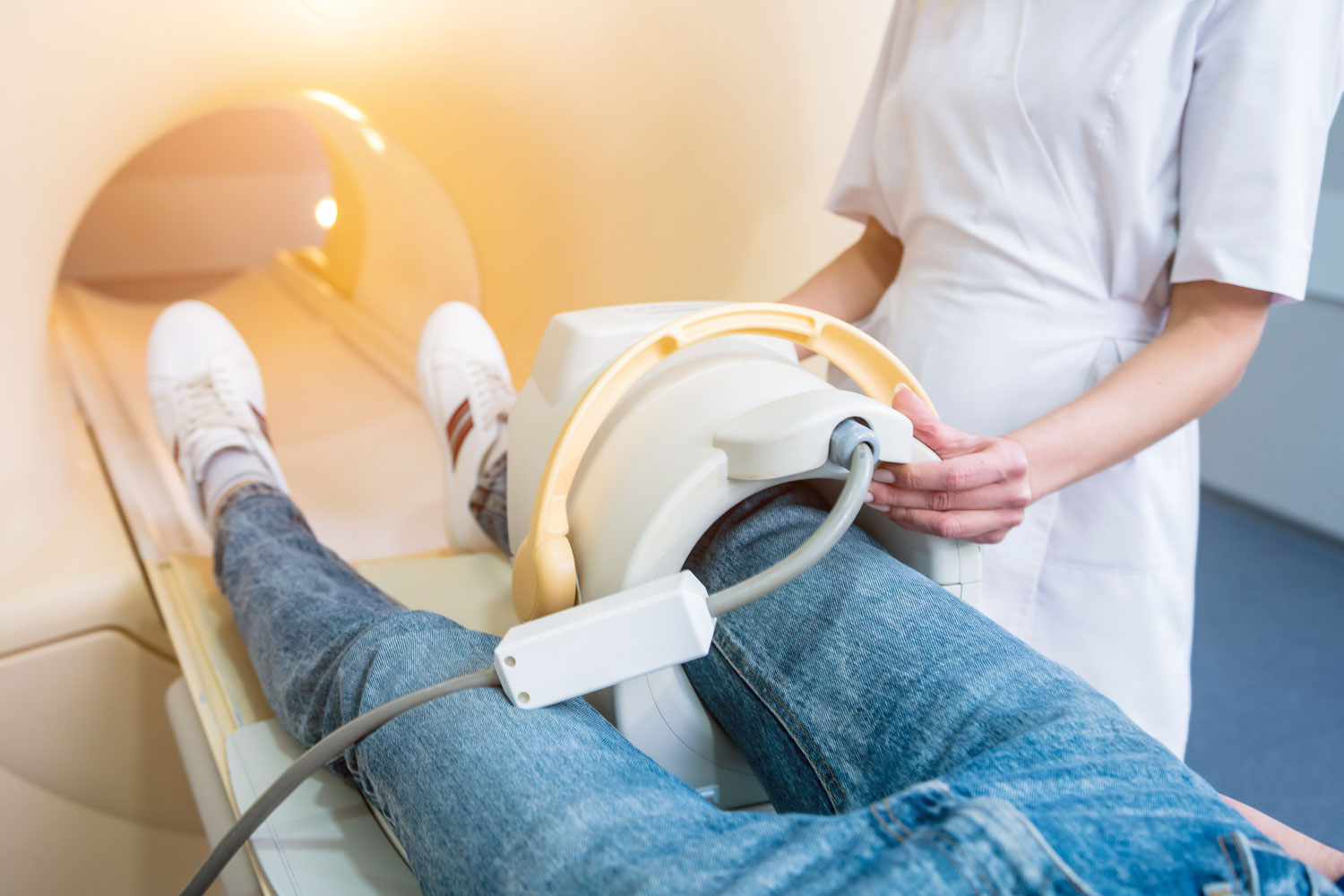
You’ll lie on a padded table and slide into the MRI machine feet first. Typically, only your lower body enters the scanner. A small coil is placed around your knee to capture the clearest images. The scan usually takes 30 to 45 minutes.
You’ll hear rhythmic tapping and humming sounds, and we’ll provide ear protection. A technologist will monitor the scan and communicate with you through an intercom. It’s important to keep your leg still throughout the scan.
In most cases, contrast dye is not needed for a standard knee MRI. However, if your doctor suspects a tumor, infection, or needs to evaluate a complex joint problem, contrast may be injected through an IV or directly into the knee joint (MR arthrography) for enhanced detail.

Magnetic resonance (MRI) of right knee, Knee joint effusion, sagittal view
Understanding Your Knee MRI Results
A board-certified radiologist will review every image and prepare a report. Key findings include:
Meniscus tears. The radiologist identifies tears by their type (horizontal, vertical, radial, or complex), location (which part of the meniscus is affected), and severity. This information helps your doctor determine whether the tear can heal on its own or may benefit from surgery.
Ligament injuries. Complete tears, partial tears, and sprains of the ACL, PCL, MCL, and LCL are identified. ACL tears, in particular, are one of the most common findings on knee MRI.
Cartilage damage. Thinning, softening, fissuring, or complete loss of the articular cartilage lining the joint surfaces is documented and graded.
Bone abnormalities. Bone bruises (contusions), stress fractures, and osteochondral lesions are identified — these are often invisible on X-ray.
Tendon and muscle injuries. Patellar tendon tears, quadriceps tendon tears, and other soft tissue injuries are evaluated.
Fluid collections. Joint effusions (excess fluid in the knee), Baker’s cysts, and bursitis are documented.
Arthritis. Early signs of osteoarthritis or inflammatory arthritis may be visible, including cartilage loss, bone marrow edema, and synovitis.
Follow-Up After Your Knee MRI
There is no recovery time. You can go home, drive, and resume normal activities immediately. Based on the results, your doctor may recommend physical therapy and rehabilitation, bracing or activity modification, anti-inflammatory medications or injections, arthroscopic surgery to repair a torn meniscus or reconstruct a ligament, or continued monitoring.
At IPMC, we work to get your results to your doctor quickly so you can start the right treatment without delay.


