
At Independent Physicians Medical Center (IPMC) in Northeast Philadelphia, we provide advanced digital chest X-ray imaging using the Siemens Multix system — a state-of-the-art digital radiography platform that delivers excellent image quality with minimal radiation in a comfortable outpatient setting. Learn more about our X-ray services.
What Does a Chest X-Ray Show?
A chest X-ray creates images by passing a controlled beam of low-dose radiation through your chest. Dense structures like bone and the heart absorb most of the radiation and appear white; air-filled lungs absorb very little and appear dark; soft tissues and fluids fall in between, showing as shades of gray. This contrast allows your radiologist to evaluate the following in a single study:
- Lungs — including the lung fields for consolidation, fluid, collapse, masses, nodules, and signs of infection or disease
- Heart — size, shape, and position; an enlarged heart (cardiomegaly) may indicate heart failure or underlying cardiac disease
- Major blood vessels — the aorta and pulmonary vessels can be evaluated for widening or abnormality
- Ribs and bones — rib fractures, bone lesions, or deformities of the chest wall
- Diaphragm — elevation, flattening (as in COPD), or free air beneath it (suggesting bowel perforation)
- Pleural space — fluid around the lungs (pleural effusion) or air (pneumothorax)
- Mediastinum — the space between the lungs, including the trachea, lymph nodes, and major vessels
Common Conditions a Chest X-Ray Detects
Pneumonia. One of the most frequent reasons a chest X-ray is ordered. Pneumonia causes the air spaces in the lungs to fill with fluid or pus, producing areas of opacity called consolidation or infiltrates. Bacterial pneumonia typically appears as a dense, well-defined opacity confined to a single lobe (lobar consolidation). Viral and atypical pneumonia (such as walking pneumonia caused by Mycoplasma) tend to produce a more diffuse, hazy pattern throughout both lungs. Bronchopneumonia produces patchy, bilateral infiltrates. A chest X-ray helps confirm the diagnosis, identify which lobe is affected, and detect complications such as pleural effusion or abscess.
Congestive heart failure and pulmonary edema. When the heart cannot pump efficiently, fluid backs up into the lungs — a condition called pulmonary edema. On a chest X-ray, this appears as haziness in the lung fields, often in a symmetrical “butterfly” or perihilar pattern, alongside an enlarged cardiac silhouette, redistribution of blood flow to the upper lobes, and Kerley B lines (fine horizontal lines at the lung bases from engorged lymphatics). A chest X-ray is often the first test used to confirm suspected heart failure.
COPD and emphysema. Chronic obstructive pulmonary disease causes the lungs to over-inflate. On X-ray, this appears as flattened diaphragms, a barrel chest configuration (increased front-to-back diameter of the thorax), and hyperlucent (darker than normal) lung fields from air trapping. Bullae — large air spaces from destroyed alveoli — may also be visible.
Pneumothorax. A collapsed lung occurs when air leaks into the pleural space between the lung and the chest wall. On a chest X-ray, a pneumothorax appears as a visible pleural line separating the deflated lung from the chest wall, with an absence of lung markings in the outer zone. A tension pneumothorax — a medical emergency — may also show a shift of the heart and mediastinum to the opposite side.
Pleural effusion. Fluid accumulation around the lung causes blunting of the costophrenic angles (the sharp corners normally seen at the base of each lung) and, when large, a homogeneous opacity at the lung base. Pleural effusions have many causes, including heart failure, pneumonia, cancer, and inflammation.
Rib fractures. Fractures of the ribs appear as breaks in the cortex of the bone on a chest X-ray, although hairline fractures and those in cartilage can be missed and may require CT for definitive evaluation.
Lung masses and nodules. Abnormal growths in the lung — ranging from benign granulomas to lung cancer — appear as white rounded densities against the darker lung background. A chest X-ray can identify larger masses; small nodules may require CT scan for further characterization and follow-up.
Tuberculosis. Active pulmonary TB classically appears as upper-lobe infiltrates, often with cavitation (air-filled holes within the consolidation). Old healed TB may leave calcified nodules or lymph nodes visible on X-ray.
Cardiomegaly. Heart enlargement is assessed by measuring the cardiothoracic ratio — the width of the heart relative to the width of the chest. A ratio greater than 50% on a PA view suggests cardiomegaly and may indicate heart failure, cardiomyopathy, or significant valvular disease.
When Does a Doctor Order a Chest X-Ray?
Chest X-rays are ordered for a wide range of clinical situations. The most common include:
- Cough — persistent, new-onset, or worsening, especially with fever or shortness of breath
- Shortness of breath or difficulty breathing
- Chest pain — to evaluate for rib fractures, pneumothorax, or cardiac causes
- Fever with respiratory symptoms — to check for pneumonia
- Suspected heart failure — to assess heart size and pulmonary congestion
- Monitoring known lung or heart conditions — such as COPD, heart failure, or pleural effusion
- Pre-operative assessment — to rule out significant cardiac or pulmonary disease before surgery
- Screening in patients with known cancer — to check for lung metastases or lymph node involvement
- Routine wellness screening — particularly in certain occupational settings or as part of an immigration or insurance medical exam
- Follow-up after treatment — to confirm resolution of pneumonia or improvement in heart failure
According to RadiologyInfo.org (published by the American College of Radiology and the Radiological Society of North America), chest X-rays are among the safest and most useful diagnostic tests available, and the radiation dose is very low — equivalent to a few days of natural background radiation.
Types of Chest X-Ray Views
Most standard chest X-rays consist of two views taken from different angles, which together give the radiologist a more complete picture of the chest anatomy.
The PA (posterior-anterior) view is the standard frontal view. You stand in front of the detector panel and the X-ray beam passes from your back to your front. This gives the truest image of heart size and lung fields and is the most commonly ordered view.
The lateral view is taken from the side — you stand with your side against the detector and the beam passes through. The lateral view is particularly helpful for identifying lesions that may be hidden behind the heart or spine on the frontal view, and for evaluating the position of lesions in relation to the lung lobes.
In some situations, additional views may be ordered. An AP (anterior-posterior) view is taken with the patient lying down and is used when a patient cannot stand — often in emergency or inpatient settings, though it slightly magnifies the heart and should not be used for cardiac size assessment. A lateral decubitus view (patient lying on their side) helps confirm a pleural effusion and distinguish free fluid from a fixed opacity.

A standard PA (posterior-anterior) chest X-ray shows the lungs as dark fields on either side of the white cardiac silhouette, with the ribs and diaphragm clearly visible. IPMC’s digital system captures this in seconds with minimal radiation.
How to Prepare for a Chest X-Ray
A chest X-ray requires virtually no preparation. You can eat, drink, and take your medications as normal. There are no fasting requirements and no restrictions beforehand. For more detail, see IPMC’s full X-ray preparation guide.
When you arrive, you will be asked to remove jewelry, necklaces, and any metal accessories from your chest and neck, as these can block X-ray penetration and obscure the image. You may be asked to change into a gown. Leave metal clothing accessories — like underwire bras — at home or be prepared to remove them.
Pregnancy: always inform the technologist if you are or may be pregnant. When a chest X-ray is medically necessary during pregnancy, lead shielding is used to protect the abdomen, and the radiation dose to a fetus from a properly shielded chest X-ray is extremely low.
What Happens During a Chest X-Ray at IPMC?
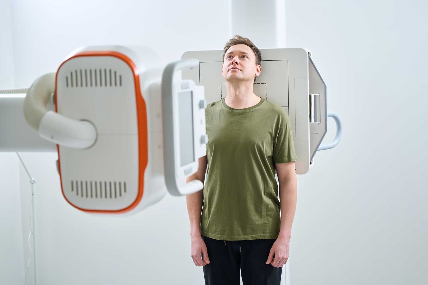
You will stand in front of the Siemens Multix digital detector panel. The technologist positions you with your chest against the panel and your chin resting on top of it. For the lateral view, you will turn 90 degrees with one shoulder closest to the panel and your arms raised or folded in front of you.
For the PA view, you will be asked to take a deep breath and hold it briefly — filling the lungs to their maximum helps expand the lung fields and improves the visibility of abnormalities. The actual exposure lasts a fraction of a second. For most standard two-view chest X-rays, the entire exam takes under 10 minutes.
There is no recovery time. You can eat, drive, and return to all normal activities immediately after the exam.
After Your Chest X-Ray: Results and Next Steps
After your exam, a board-certified radiologist at IPMC reviews every image and prepares a detailed written report for your referring physician. Results are typically available within 24 to 48 hours for outpatient studies and sent directly to your doctor.
A normal chest X-ray shows clear lung fields, a heart of normal size, sharply defined ribs and diaphragm, and no visible masses, nodules, or fluid accumulation.
An abnormal chest X-ray may show one or more findings that prompt your doctor to recommend next steps — which could include a course of antibiotics for pneumonia, a repeat X-ray in follow-up, a CT scan of the chest for more detailed evaluation of a nodule or mass, referral to a pulmonologist or cardiologist, or other targeted workup depending on the specific finding.
Not all abnormal findings on a chest X-ray indicate serious disease — many represent old changes, minor incidental findings, or findings that require only monitoring. Your referring physician will explain what the specific findings mean for your clinical situation.
Chest X-Ray vs. Chest CT: When Is More Detail Needed?
A chest X-ray is fast, low-dose, and excellent for detecting large-scale abnormalities — but its resolution has limits. When a chest X-ray identifies a finding that needs more detailed characterization, or when the index of suspicion is high despite a normal X-ray, a CT scan of the chest is typically the next step. CT provides cross-sectional images with far greater detail — able to detect small nodules, characterize masses, evaluate the airways, and assess the mediastinum and pleura in ways a plain film cannot.
For cardiac evaluation beyond heart size, an MRI or echocardiogram provides detailed structural and functional assessment of the heart without radiation. See our full CT scan vs. MRI comparison guide for more on choosing between imaging modalities.


