
A cervical MRI scan gives your doctor an unmatched view of these vertebrae, discs, nerves, and surrounding soft tissues. If you’re searching for a cervical MRI in Philadelphia for neck pain, a pinched nerve, or upper spine problems, IPMC provides advanced imaging in a comfortable outpatient setting in Northeast Philadelphia.
What Is a Cervical MRI?
A cervical MRI is a noninvasive imaging test that uses magnetic fields and radio waves to create detailed pictures of the seven cervical vertebrae, the intervertebral discs between them, the spinal cord as it passes through the neck, the nerve roots that branch off to the arms and hands, and all of the ligaments, muscles, and blood vessels in the area.
This test is the gold standard for examining soft tissue structures in the neck because it provides far more detail than X-rays or CT scans, all without ionizing radiation. It can reveal problems that are completely invisible on other types of imaging.
Why Would Your Doctor Order a Cervical MRI?
Your physician may recommend a cervical MRI if you are experiencing:
- Persistent neck pain or stiffness that hasn’t responded to conservative treatment over several weeks
- Pain, numbness, tingling, or a “pins and needles” sensation that radiates from the neck into one or both shoulders, arms, or hands — this pattern often indicates a compressed nerve root (cervical radiculopathy)
- Weakness in your arms, hands, or grip strength
- Difficulty with fine motor tasks such as buttoning a shirt, writing, or picking up small objects
- Problems with balance, coordination, or walking — which may suggest spinal cord compression (cervical myelopathy)
- Suspected herniated or bulging disc in the neck
- Signs of cervical spinal stenosis (narrowing of the spinal canal)
- Neck injury or trauma, such as whiplash from a car accident or a sports injury
- Suspected multiple sclerosis — MRI can detect characteristic lesions (plaques) in the spinal cord
- Evaluation of tumors, infections, or inflammatory conditions affecting the cervical spine
- Pre-surgical planning for cervical spine procedures such as discectomy, fusion, or artificial disc replacement
- Post-surgical follow-up to evaluate healing and check for complications
A cervical MRI can detect herniated discs, bone spurs compressing nerves, spinal cord compression, spinal stenosis, degenerative disc disease, fractures, tumors, infections, demyelinating diseases like MS, and vascular abnormalities.
How to Prepare for Your Cervical MRI
For most cervical MRIs, you can eat, drink, and take your medications as usual. Remove all metal objects including necklaces, earrings, and hair accessories. Inform your care team about any metal implants, especially if you’ve had prior cervical spine surgery with hardware. Wear comfortable clothing without metal. If you’re claustrophobic, tell your doctor in advance — for a cervical MRI, your head and neck will be inside the scanner, so accommodations may be helpful.
What Happens During the Scan?
You’ll lie on a cushioned table, and a special coil will be placed around your neck to capture the clearest images. The table slides into the MRI machine. The scan typically takes 30 to 60 minutes. You’ll hear tapping and humming noises and will receive ear protection. A technologist communicates with you through an intercom at all times. It’s important to stay as still as possible. If contrast dye is needed, it will be administered through an IV.

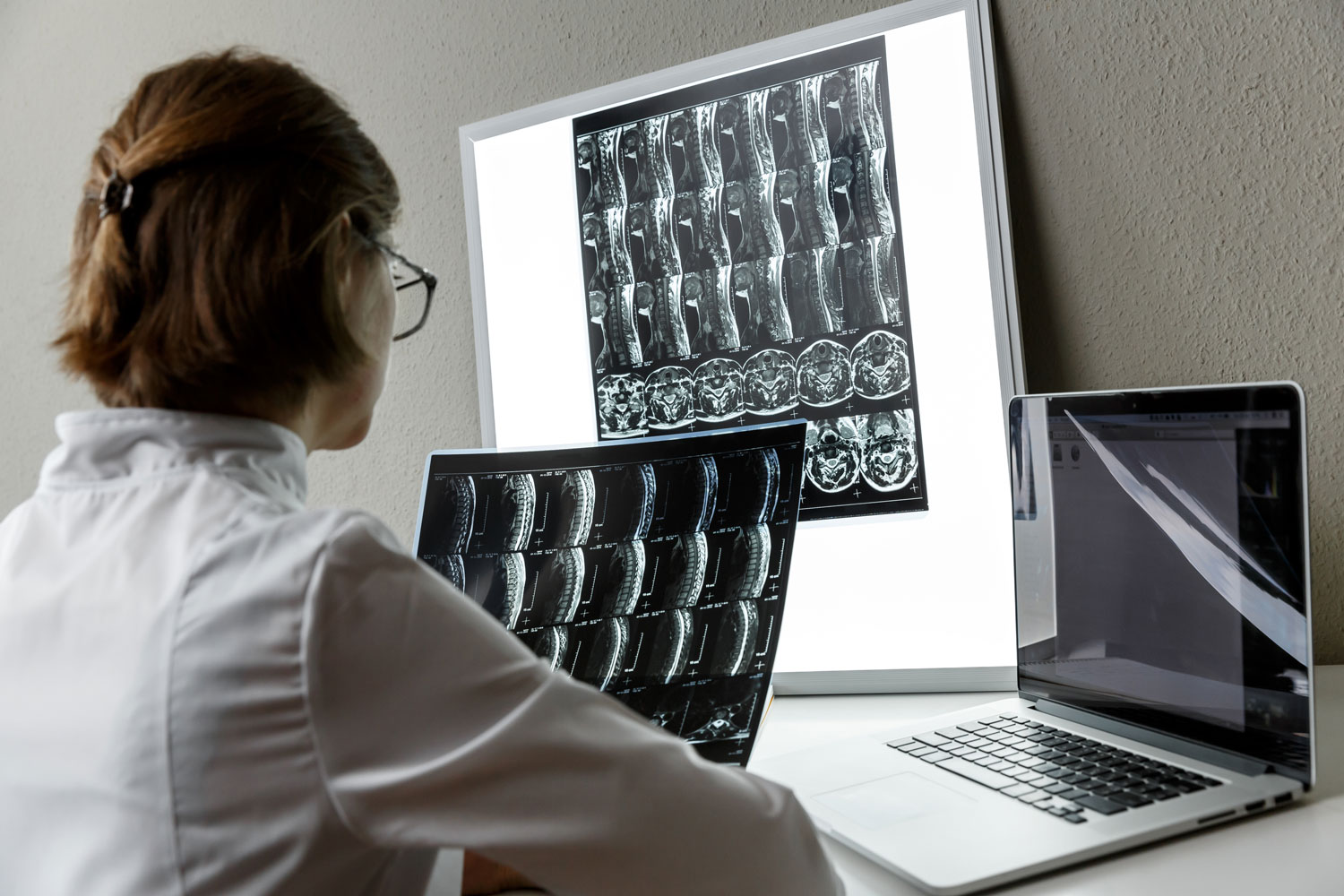
Cervical MRI scan showing spine with and without contrast
Understanding Your Cervical MRI Results
A board-certified radiologist at IPMC will carefully analyze every image and prepare a detailed report for your referring physician. Key findings include:
Disc herniations and bulges. The radiologist identifies whether any cervical discs have protruded beyond their normal position and whether displaced disc material is pressing on a nerve root or the spinal cord. The level (e.g., C5-C6 or C6-C7), direction, and severity are all documented.
Nerve root compression (radiculopathy). Narrowing of the neural foramina — the openings where nerve roots exit the spine — is identified and graded. This helps explain symptoms like arm pain, numbness, or weakness.
Spinal cord compression (myelopathy). Any compression or signal changes within the spinal cord itself are noted. Cord signal changes can indicate damage from long-standing compression and influence the urgency of treatment.
Cervical stenosis. Narrowing of the spinal canal from disc bulges, bone spurs, or thickened ligaments is measured and graded as mild, moderate, or severe.
Degenerative changes. Disc dehydration, bone spurs (osteophytes), facet joint arthritis, and endplate changes are documented as part of the overall assessment of cervical spine health.
Tumors, infections, and demyelinating disease. Masses, signs of infection, or MS-related lesions in the spinal cord are identified and characterized.
Follow-Up After Your Cervical MRI
There is no recovery time after a cervical MRI. You can return to your normal activities, diet, and medications right away.
Based on the findings, your doctor may recommend physical therapy and targeted exercises for neck strengthening, oral medications or nerve pain treatments, cervical epidural steroid injections to relieve nerve compression symptoms, referral to a spine specialist, neurologist, or neurosurgeon, or surgical options such as anterior cervical discectomy and fusion (ACDF) or artificial disc replacement for severe cases.
At IPMC, we work to deliver results to your doctor promptly — so your treatment plan can begin without unnecessary delays.fmult


