
When kidney stones are suspected or known, imaging is essential — to confirm the diagnosis, identify the stone’s location, assess whether it is causing obstruction, and guide treatment decisions. A standard abdominal X-ray — specifically a KUB X-ray (kidneys, ureters, and bladder) — is the simplest, fastest, and lowest-radiation option available. It can detect the most common types of kidney stones quickly and inexpensively. But it has real limitations, and knowing what it can and cannot show helps both patients and doctors use it appropriately.
At Independent Physicians Medical Center (IPMC) in Northeast Philadelphia, we offer both KUB X-ray and CT imaging at the same convenient outpatient facility — so your doctor can order whichever study is most appropriate, or both, without sending you to multiple locations. Learn more about our X-ray services.
What Is a KUB X-Ray?
A KUB is a single anteroposterior (AP) plain X-ray of the abdomen taken with the patient lying flat on their back. The image covers the entire abdomen from the upper kidney poles to the bladder — encompassing the full course of both ureters. It is the same basic X-ray technology used for other abdominal studies, with no contrast agent, no preparation, and no radiation beyond that of a standard abdominal film.
On a KUB, dense calcium-containing structures appear white — and this is the principle on which kidney stone detection is based. A calcium stone bright against the background of soft tissue is visible as a small, dense white opacity overlying the kidney, ureter, or bladder. The study takes just a few minutes from positioning to completion, making it one of the fastest imaging tests available.
Which Kidney Stones Show Up on X-Ray?
Whether a kidney stone is visible on X-ray depends entirely on its composition. Stones that contain calcium absorb X-rays and appear white; stones that do not contain calcium are radiolucent and completely invisible.
Calcium oxalate and calcium phosphate stones make up approximately 75–80% of all kidney stones and are strongly radiopaque — they typically show up clearly as bright white densities on the KUB. These are the stones a plain X-ray is best at detecting.
Struvite stones (also called infection stones or triple-phosphate stones) account for about 10–15% of stones and form in the setting of urinary tract infections with urea-splitting bacteria. They are moderately radiopaque and are often visible on X-ray, particularly when large. Struvite stones can grow to fill the entire renal collecting system — producing the characteristic “staghorn calculus” shape that is readily visible on plain film.
Uric acid stones account for 5–10% of kidney stones and are entirely radiolucent — they contain no calcium and are invisible on KUB X-ray. Uric acid stones are more common in patients with gout, diabetes, obesity, and those who produce highly acidic urine. The distinction matters clinically: uric acid stones can often be dissolved with urinary alkalinization (no surgery needed), so identifying them correctly guides treatment. A stone that is visible on CT but invisible on KUB is often presumed to be uric acid.
Cystine stones are moderately radiopaque — they contain sulfur, which provides some density. They are typically visible on X-ray but less bright than calcium stones.
Very small stones of any composition may be missed on KUB, even calcium-containing ones. Stones under 2–3 mm are often below the resolution of plain X-ray and can be obscured by overlying bowel gas, stool, or bony structures.
The Limitations of X-Ray for Kidney Stone Diagnosis
A KUB X-ray is a useful tool in kidney stone management, but it should not be the sole imaging study relied upon when definitive diagnosis is needed. Its limitations are significant and clinically important.
It misses radiolucent stones entirely. Uric acid stones, certain mixed-composition stones, and drug-induced stones (such as indinavir stones in HIV patients) are completely invisible on KUB. A normal KUB does not rule out kidney stone disease if the stone is not calcium-based.
It cannot assess obstruction. Even when a stone is visible on X-ray, the KUB cannot tell whether the stone is causing obstruction of the ureter — a critical clinical question. An obstructing stone that is preventing urine from draining can cause progressive kidney damage and, if complicated by infection, becomes a urological emergency. Only CT (or less commonly, ultrasound or intravenous pyelogram) can assess whether the kidney is obstructed.
Overlapping structures cause false positives and false negatives. Phleboliths (calcified veins in the pelvis), calcified lymph nodes, gallstones, costal cartilage calcifications, and other incidental densities can mimic kidney stones on KUB — producing false-positive appearances. Conversely, real stones can be hidden by overlapping bowel gas, stool, or bony structures. The overall sensitivity of KUB for kidney stones is approximately 44–77% in the literature — significantly lower than the 95%+ of non-contrast CT.
It cannot characterize the urinary tract anatomy. The KUB shows stones but not the surrounding urinary tract structures. CT provides detailed anatomy of the kidneys, ureters, and bladder — important for assessing conditions that may mimic stones (such as a ureteropelvic junction obstruction) or that coexist with them.
When Is a KUB X-Ray the Right Choice?
Despite its limitations, the KUB has a well-defined and valuable role in kidney stone management. It is not the right test for every situation, but for the right patients it is fast, affordable, and low-radiation.
Monitoring a known calcium stone. Once a calcium stone has been identified on CT, follow-up KUB X-rays can track its position and size over time without the significantly higher radiation exposure of repeated CT scans. This is one of the most common uses of KUB — serial monitoring during watchful waiting or medical expulsive therapy.
Confirming stone passage after treatment. After lithotripsy (ESWL — extracorporeal shock wave lithotripsy), ureteroscopy, or spontaneous stone passage, a KUB can confirm that the stone is no longer present or has fragmented into smaller passable pieces — without the radiation burden of CT.
Pre-operative planning by the urologist. Urologists often obtain a KUB before shock wave lithotripsy or surgical stone removal to confirm the stone’s current position and assess whether it is still accessible by the intended approach.
Screening in recurrent stone formers. Patients who form calcium stones repeatedly may undergo periodic KUB studies to check for new stone formation in the kidney — a cost-effective monitoring strategy compared to CT.
Initial quick assessment in selected low-risk patients. In a stable patient with a known history of calcium stones who presents with mild symptoms consistent with their usual stone episodes, a KUB can provide a rapid initial look while planning further evaluation.
When Is CT Scan the Right Choice?
A non-contrast CT scan of the abdomen and pelvis is the gold standard for kidney stone evaluation. With sensitivity exceeding 95% for stones of all compositions, it identifies the stone’s exact location, size, and composition characteristics; shows whether the ureter is obstructed; reveals the degree of hydronephrosis (kidney swelling from blocked outflow); and can detect alternative diagnoses — such as appendicitis, ovarian pathology, aortic aneurysm, or other causes of acute flank pain — that can mimic kidney stones clinically.
CT is the appropriate choice when:
- The diagnosis is uncertain and a definitive answer is needed
- Obstruction needs to be assessed — especially with fever, suggesting an infected obstructed kidney (a urological emergency)
- The KUB is negative but stone is still clinically suspected
- It is a first presentation of flank pain and an alternative diagnosis must be excluded
- Pre-operative planning requires precise stone location, size, and density
- The stone composition is unknown or uric acid stones are suspected
IPMC offers CT imaging at the same facility as KUB X-ray — if your doctor needs both tests, you can complete them in a single visit.
How to Prepare and What to Expect
A KUB X-ray requires no preparation. You can eat, drink, and take your medications as normal. For complete preparation guidelines, see IPMC’s X-ray preparation page.
If you have recently had a CT scan with contrast dye or a barium study, inform your scheduling team — residual contrast material in the abdomen can obscure kidney stones on a plain KUB film, and your doctor may recommend waiting 10 days before the X-ray.
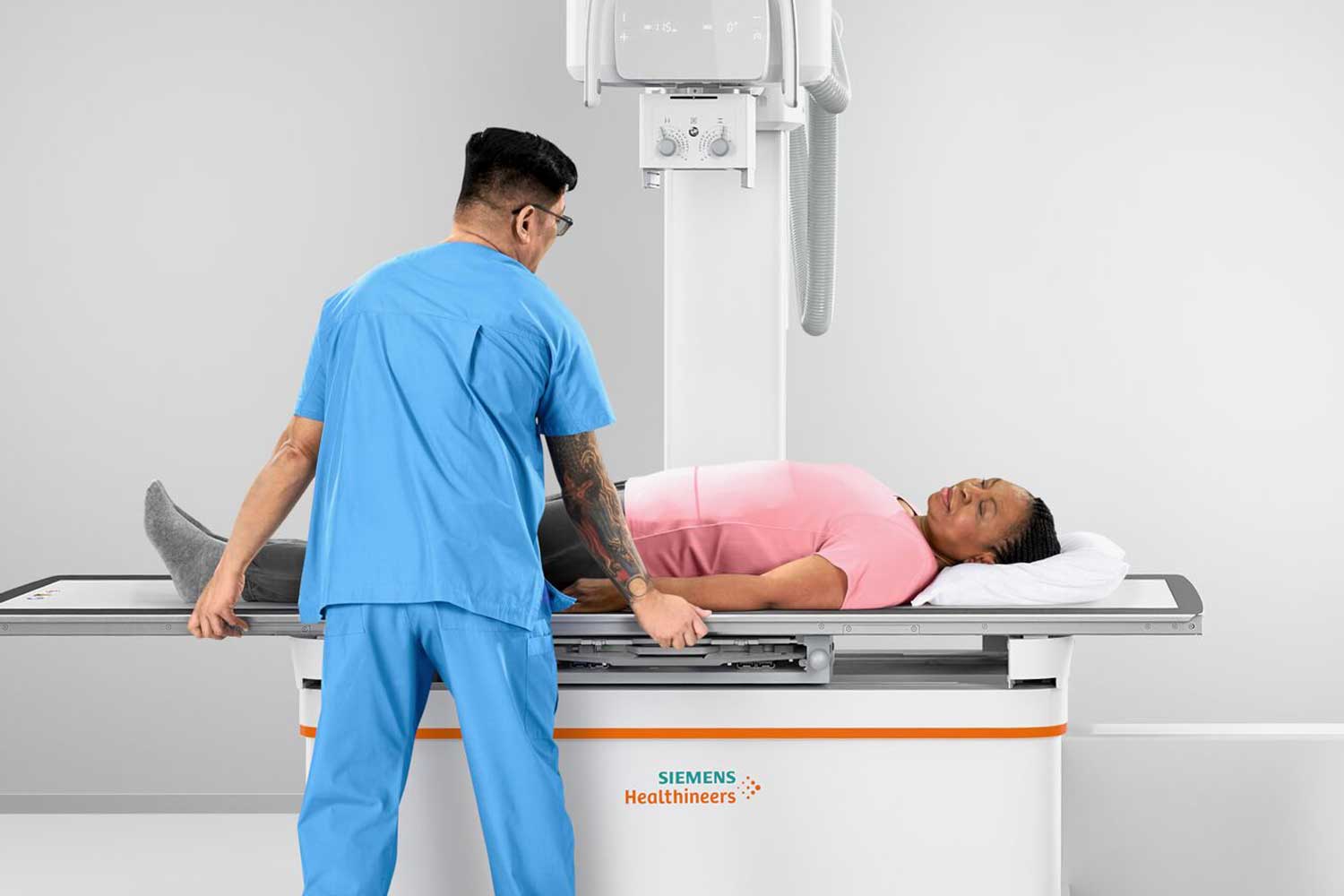
You will lie flat on the X-ray table. The technologist takes a single frontal view covering the entire abdomen from the kidneys to the bladder. The exam takes just a few minutes and is completely painless — there are no injections, no contrast, and no recovery time. You can drive and return to all normal activities immediately after.
Pregnancy: always inform the technologist if you are or may be pregnant. The KUB directs the X-ray beam directly through the abdomen and pelvis. When imaging is medically necessary during pregnancy, the clinical benefit is weighed carefully, and ultrasound or MRI (no radiation) may be preferred for initial kidney stone evaluation in pregnant patients.
After Your KUB X-Ray: Results and Next Steps
A board-certified radiologist at IPMC reviews every image and prepares a detailed written report for your referring physician, typically within 24 to 48 hours. Your doctor will interpret the findings in the context of your symptoms, urinalysis, and clinical history.
If a stone is visible, the report will describe its location (kidney, ureter, or bladder), approximate size, and density. Your urologist or primary care doctor will use this to guide management — watchful waiting, medical expulsive therapy, or intervention.
If no stone is visible, the KUB result should be interpreted carefully. A negative KUB does not rule out kidney stones — it rules out large calcium stones in easily visible locations. If your symptoms strongly suggest a stone, a CT scan of the abdomen and pelvis is the next step for definitive evaluation.
If a density is seen but its nature is uncertain, your doctor may recommend a CT scan to confirm whether it is truly a urinary stone or an incidental finding such as a phlebolith or calcified lymph node.
For more on understanding imaging results in the context of your symptoms, ask your urologist or referring physician — IPMC’s results are sent directly to your doctor, who will discuss the findings and next steps with you.
X-rays at IPMC
Why Choose IPMC for Your Kidney Stone Imaging in Philadelphia?
KUB X-Ray and CT Scan at the Same Location
IPMC offers both KUB X-ray and non-contrast CT at the same convenient Northeast Philadelphia location. If your doctor needs both — or needs to upgrade from X-ray to CT in the same visit — you can complete everything without traveling to a second facility.
Board-Certified Radiologists
Every KUB X-ray is interpreted by a board-certified radiologist experienced in abdominal and urological imaging. Reports — describing stone location, size, and recommendations for follow-up — are sent directly to your referring physician within 24 to 48 hours.
Fast Appointments, No Hospital Wait Times
A KUB X-ray at IPMC takes just a few minutes. Same-day and next-day scheduling is often available. When you need quick answers about kidney stone progression or passage, you don’t have to wait days for a hospital appointment.
Convenient Location and Flexible Hours
Located at 9908 E. Roosevelt Blvd. in Northeast Philadelphia with onsite parking. Open Monday–Friday, 8AM–8PM. We accept most major insurance plans.
Schedule Your Kidney Stone Imaging at IPMC in Philadelphia
If your doctor has recommended a KUB X-ray or CT scan for kidney stones in Philadelphia, IPMC provides fast, expert imaging in a comfortable outpatient setting in Northeast Philadelphia — with results delivered directly to your physician.
- Call 215-464-3300 to schedule your appointment.
- 9908 E. Roosevelt Blvd., Philadelphia, PA 19115
- Monday–Friday, 8AM–8PM


