
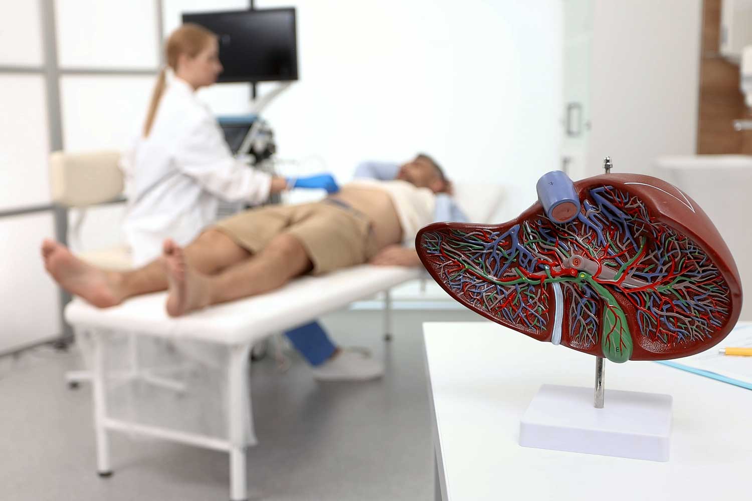
At Independent Physicians Medical Center (IPMC) in Northeast Philadelphia, we provide liver ultrasound imaging and can help your doctor evaluate your liver health with clarity and care.
What Does a “Normal” Liver Look Like on Ultrasound?
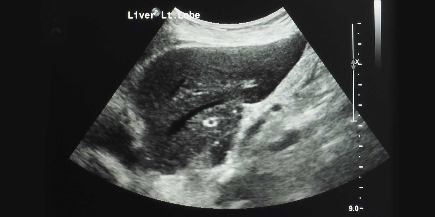
On a normal liver ultrasound, the liver appears as a smooth-bordered organ with uniform echogenicity (consistent brightness) that is similar to or slightly brighter than the adjacent kidney. The liver’s internal blood vessels (portal veins and hepatic veins) are clearly visible, and the bile ducts are thin and not dilated. The liver is a normal size (the right lobe typically measures less than 15-16 cm in length).
When any of these characteristics deviate from normal, the radiologist will describe the finding as abnormal and provide details about what was seen.
Common Abnormal Findings on Liver Ultrasound
Here are some of the most common abnormal findings and what they may mean:
- Fatty liver (hepatic steatosis). This is the most common abnormal finding on liver ultrasound. The liver appears brighter (more echogenic) than normal, indicating fat accumulation within the liver cells. Fatty liver disease can be caused by obesity, diabetes, high cholesterol, alcohol use, or certain medications. Mild fatty liver is very common and often reversible with lifestyle changes. However, in some cases, it can progress to inflammation (steatohepatitis) or scarring (fibrosis)
- Liver cysts. Simple cysts are fluid-filled sacs that are extremely common, especially with aging, and are almost always benign. They appear as dark, round, well-defined structures on ultrasound. They rarely cause symptoms or require treatment
- These are the most common benign liver tumors. They are made up of a tangle of blood vessels and appear as well-defined, bright (echogenic) masses on ultrasound. They are almost always harmless and are typically monitored rather than treated
- Liver masses or nodules. Solid masses in the liver can be benign or malignant. Ultrasound can detect them, but further characterization with CT or MRI is usually needed to determine their nature. Common benign masses include focal nodular hyperplasia (FNH) and hepatic adenomas. Malignant masses include hepatocellular carcinoma (primary liver cancer) and metastases from cancers elsewhere in the body
- Advanced scarring of the liver changes its appearance on ultrasound — the surface becomes nodular and irregular, the liver may be shrunken, and the spleen may be enlarged. The portal vein may show signs of portal hypertension
- Bile duct dilation. Widening of the intrahepatic or common bile ducts suggests an obstruction, which may be caused by gallstones, strictures, or tumors
- Hepatomegaly (enlarged liver). An enlarged liver can result from fatty liver disease, hepatitis, heart failure, infections, or infiltrative conditions
- Free fluid in the abdomen surrounding the liver may indicate liver disease, heart failure, infection, or cancer
- Abnormal blood flow. Doppler ultrasound may show abnormal flow in the portal vein (portal hypertension, portal vein thrombosis) or hepatic veins (Budd-Chiari syndrome)
What Happens After an Abnormal Liver Ultrasound?
Your doctor will consider your ultrasound findings alongside your symptoms, physical exam, and blood test results to determine the best next step. Depending on the finding, this may include:
Blood tests to assess liver function (ALT, AST, alkaline phosphatase, bilirubin, albumin) and look for specific causes of liver disease (hepatitis panel, iron studies, autoimmune markers).
Additional imaging such as CT scan or MRI for more detailed characterization of masses, nodules, or other findings that need further evaluation. MRI with contrast is often the best test for characterizing liver lesions.
Elastography (FibroScan or MR elastography) to measure liver stiffness if fibrosis or cirrhosis is suspected.
Follow-up ultrasound at regular intervals to monitor stable findings like small cysts, hemangiomas, or mild fatty liver.
Lifestyle modifications for fatty liver disease — weight loss, dietary changes, exercise, and reducing alcohol intake can significantly improve or reverse fatty liver.
Referral to a hepatologist (liver specialist) or gastroenterologist for complex or concerning findings.
The most important step is to follow up with your doctor so that any issues can be addressed early, when treatment is most effective.


