
At Independent Physicians Medical Center (IPMC) in Northeast Philadelphia, we offer carotid duplex ultrasound imaging to help your doctor assess your vascular health and stroke risk.
What Is a Carotid Duplex Ultrasound?
A carotid duplex ultrasound combines two types of imaging into one exam:
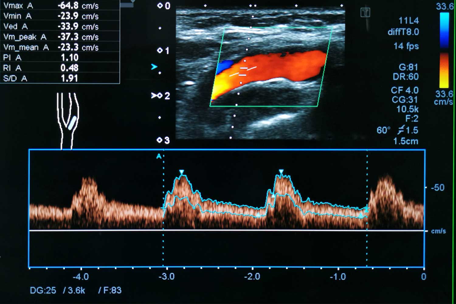
B-mode (brightness mode) ultrasound produces grayscale images that show the structure of the carotid arteries, including the vessel walls, any plaque buildup, and the presence of narrowing. It can also characterize plaque as stable (calcified, smooth) or potentially unstable (soft, irregular, ulcerated) — unstable plaque carries a higher stroke risk.
Doppler ultrasound measures the speed and direction of blood flow through the carotid arteries. When an artery is narrowed, blood speeds up as it passes through the tight area — similar to how water flows faster through a partially closed garden hose. By measuring these velocities, the sonographer and radiologist can estimate the degree of narrowing (stenosis) with remarkable accuracy.
Color Doppler adds color to the image to visualize flow direction (red for flow toward the transducer, blue for flow away), while spectral Doppler produces a waveform graph of flow velocities at specific points in the artery.
Why Would Your Doctor Order a Carotid Duplex Ultrasound?
Your doctor may recommend this test if you:
- Have had a stroke or transient ischemic attack (TIA / “mini-stroke”). This is one of the most common reasons for the test. After a stroke or TIA, evaluating the carotid arteries for significant narrowing is a critical part of determining the cause and preventing future events
- Have a carotid bruit. This is an abnormal whooshing sound detected with a stethoscope when listening to the neck, which may indicate turbulent blood flow from a narrowed artery
- Have significant risk factors for stroke or cardiovascular disease, including high blood pressure, high cholesterol, diabetes, smoking, a family history of stroke, or peripheral artery disease
- Are being evaluated before heart surgery (such as coronary artery bypass grafting), as the status of the carotid arteries affects surgical risk
- Have known carotid artery disease that is being monitored over time to track progression or stability
- Had a previous carotid endarterectomy (surgery) or carotid stent placement and need follow-up to check patency
- Have unexplained dizziness, visual disturbances, or neurological symptoms that could relate to reduced blood flow to the brain
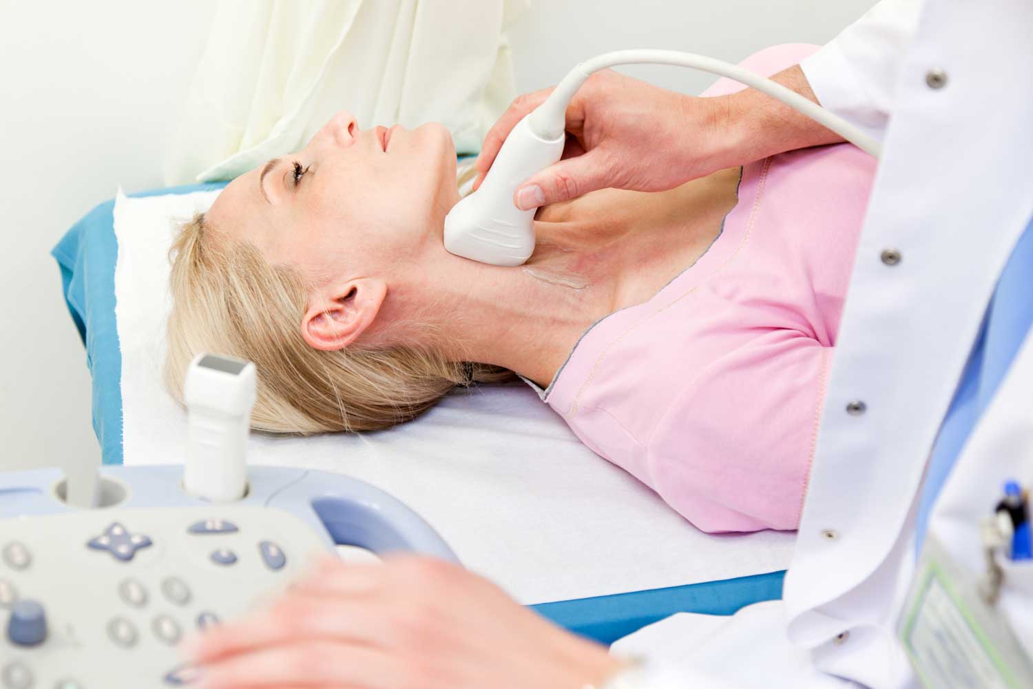
What Happens During the Exam?
No preparation is needed. You can eat, drink, and take your medications normally. Wear a shirt with an open or low collar for easy access to your neck.
You’ll lie on your back with your head turned slightly to one side. The sonographer applies gel to your neck and uses a transducer to capture images and blood flow measurements from both carotid arteries, the internal and external carotid branches, and the vertebral arteries. The sonographer systematically evaluates each vessel, measuring flow velocities and documenting any plaque or narrowing.
The exam is painless and typically takes 30 to 45 minutes. Both sides are always examined, even if symptoms are only on one side.
Understanding Your Results
A board-certified radiologist reviews the images and Doppler measurements and grades the degree of carotid stenosis using standardized velocity criteria:
Normal (no stenosis). Blood flows smoothly with normal velocities.
Mild stenosis (less than 50%). Some plaque is present but the narrowing is not hemodynamically significant. Monitoring is typically recommended.
Moderate stenosis (50-69%). More significant narrowing that may require closer monitoring or treatment depending on symptoms.
Severe stenosis (70% or greater). Significant narrowing that substantially increases stroke risk. For symptomatic patients (those who have had a stroke or TIA), surgical treatment (carotid endarterectomy or stenting) is often recommended.
Near-occlusion or total occlusion. The artery is nearly or completely blocked.
Your doctor will discuss the results and recommended action, which may include lifestyle modifications and risk factor management, medications (blood thinners, cholesterol-lowering drugs, blood pressure control), surgical intervention for severe or symptomatic stenosis, or regular follow-up ultrasounds to monitor progression.


