
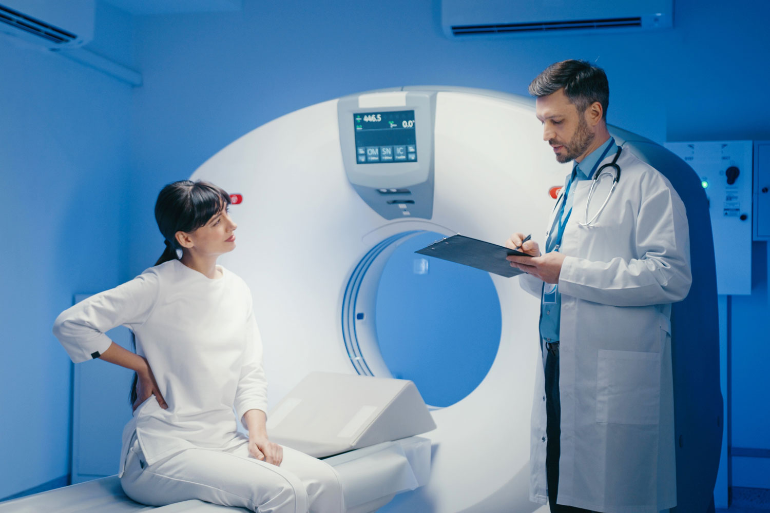
At Independent Physicians Medical Center (IPMC) in Northeast Philadelphia, we provide lumbar MRI imaging with advanced technology, experienced technologists, and board-certified radiologists in a comfortable outpatient environment.
What Is a Lumbar MRI?
A lumbar MRI uses powerful magnets and radio waves to create highly detailed cross-sectional images of your lower spine, also known as the lumbar region. This area includes the five lumbar vertebrae (L1 through L5), the sacrum (the triangular bone at the base of the spine), the coccyx (tailbone), and the surrounding structures — including intervertebral discs, the spinal cord and nerve roots, ligaments, muscles, and the spinal canal.
Unlike X-rays, which primarily show bones, a lumbar MRI provides exceptional soft tissue detail. This makes it the preferred imaging test for investigating back problems that involve discs, nerves, the spinal cord, and the small joints of the spine. Because it uses no ionizing radiation, it is safe for repeated use and for patients of all ages.
Why Would Your Doctor Order a Lumbar MRI?
Your doctor may recommend a lumbar MRI if you are experiencing:
- Persistent lower back pain that hasn’t improved after four to six weeks of conservative treatment such as rest, physical therapy, and medication
- Pain, numbness, or tingling that radiates from the lower back down into one or both legs (sciatica) — this often suggests nerve compression from a herniated disc or spinal stenosis
- Significant leg weakness, difficulty walking, or a foot drop (inability to lift the front of the foot)
- Loss of bladder or bowel control — this is a medical emergency called cauda equina syndrome that requires urgent imaging and treatment
- Back pain accompanied by fever, which may suggest a spinal infection (discitis or osteomyelitis)
- Suspected herniated or bulging disc pressing on a nerve root
- Signs of spinal stenosis — a narrowing of the spinal canal that compresses the nerves, often causing pain, numbness, or weakness in the legs, especially with walking
- Suspected spinal tumors, whether primary or metastatic (cancer that has spread from another part of the body)
- Degenerative disc disease or other age-related changes in the spine
- Pre-surgical planning for spinal procedures such as decompression, fusion, or disc replacement
- Post-surgical follow-up to evaluate healing, check for complications, or investigate new or recurrent symptoms
A lumbar MRI can detect herniated and bulging discs, spinal stenosis, nerve root compression, spondylolisthesis (forward slippage of a vertebra), fractures (including compression fractures and stress fractures), infections, tumors, cauda equina syndrome, and degenerative changes. It provides the information your doctor needs to make an accurate diagnosis and choose the most effective treatment.
How to Prepare for Your Lumbar MRI
Eating and medications. For most lumbar MRIs, you can eat, drink, and take your medications normally. If contrast dye will be used, your doctor may give specific fasting instructions.
Metal safety. Remove all metal objects and inform your care team about any implants, surgical hardware, or metal in your body. If you have spinal hardware from a previous surgery (such as rods, screws, or cages), let the technologist know — most modern spinal implants are MRI-compatible, but this should always be verified.
Clothing. Wear comfortable clothes without metal. You may change into a gown.
Contrast dye. If your doctor suspects infection, tumor, or needs to evaluate post-surgical changes, contrast dye may be ordered. Let your care team know about any allergies or kidney problems.
Claustrophobia. If enclosed spaces make you anxious, tell your doctor in advance.
What Happens During a Lumbar MRI?
At IPMC, you’ll lie on your back on a cushioned table. A coil may be placed over your lower back. The table slides into the MRI machine, and the scan typically takes 30 to 60 minutes.
During the scan, the machine produces loud tapping and humming sounds. We’ll provide ear protection and can often play music through headphones. A technologist will communicate with you via intercom throughout the entire exam. You’ll need to lie still for the clearest images, and the technologist may ask you to hold your breath briefly for certain sequences.
If contrast dye is used, it will be injected through a small IV partway through the scan, and additional images will be captured.
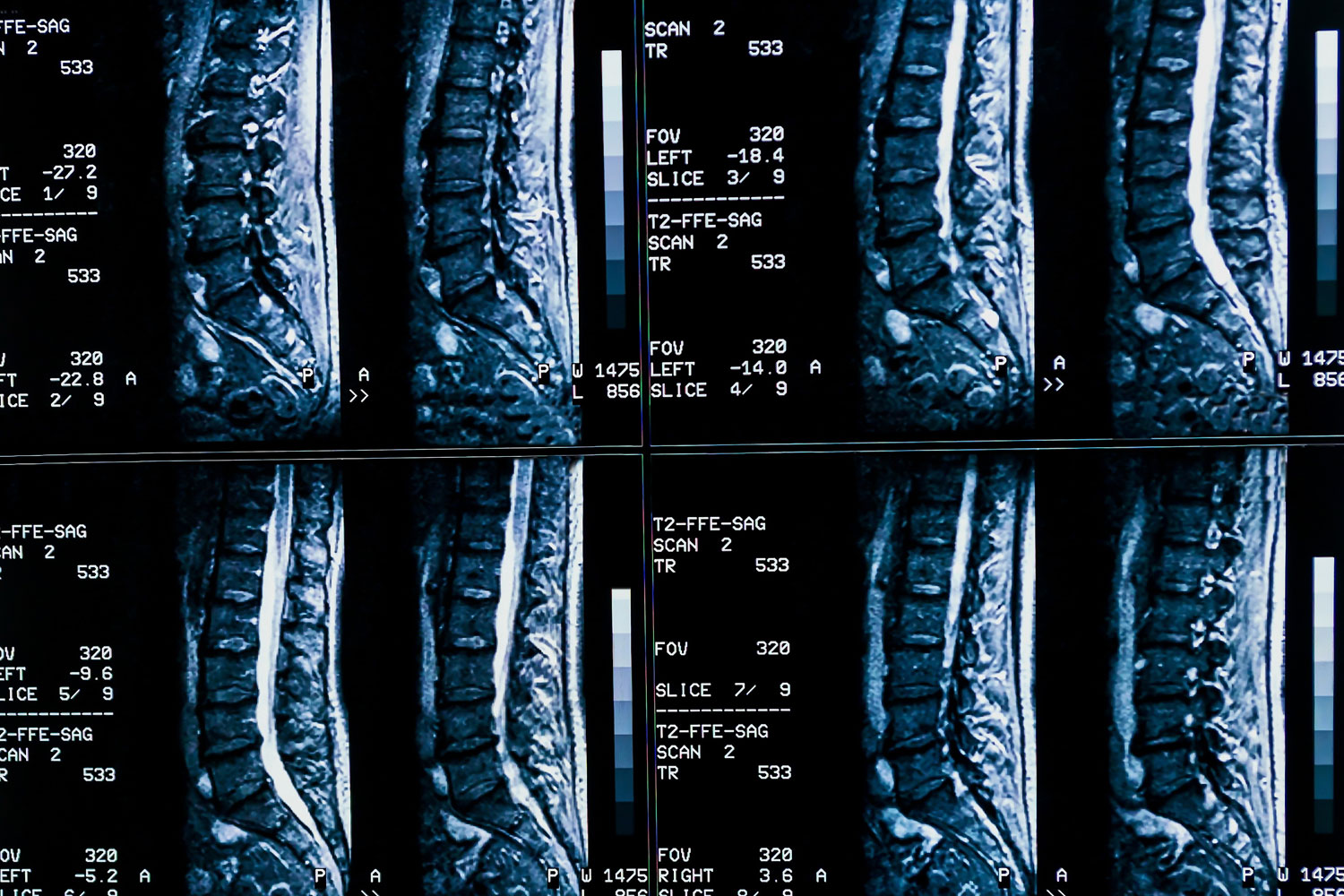
The MRI shows degenerative changes of spines, lumbar discs herniation and nerve roots compression.
Understanding Your Lumbar MRI Results
A board-certified radiologist will analyze every image and prepare a detailed report. Key findings your radiologist looks for include:
Disc herniations. The radiologist identifies whether any discs have bulged or herniated (protruded) beyond their normal position, and whether the displaced disc material is pressing on a nerve root or the spinal cord. The specific location and severity of the herniation are documented.
Spinal stenosis. Narrowing of the spinal canal or the openings where nerve roots exit the spine (foraminal stenosis) is measured and graded as mild, moderate, or severe.
Nerve compression. Any evidence that a nerve root is being compressed — by a disc, bone spur, or other structure — is identified and correlated with your symptoms.
Degenerative changes. Disc dehydration (loss of disc height and water content), facet joint arthritis, bone spurs, and endplate changes (Modic changes) are documented.
Fractures. Compression fractures, stress fractures, and traumatic fractures are identified. MRI is especially sensitive at detecting fractures that are not visible on X-rays.
Tumors, infections, and other conditions. Any masses, areas of abnormal signal, or signs of infection are identified and characterized.
Follow-Up After Your Lumbar MRI
There is no downtime after a lumbar MRI. You can return to your normal activities right away, unless you received sedation for anxiety.
Based on the findings, your doctor may recommend physical therapy and exercise, pain management with medications or injections (such as epidural steroid injections), referral to a spine specialist or neurosurgeon, continued monitoring, or surgery for severe conditions that haven’t responded to conservative treatment.
At IPMC, we deliver results to your doctor as quickly as possible so your treatment plan can move forward without unnecessary waiting.


